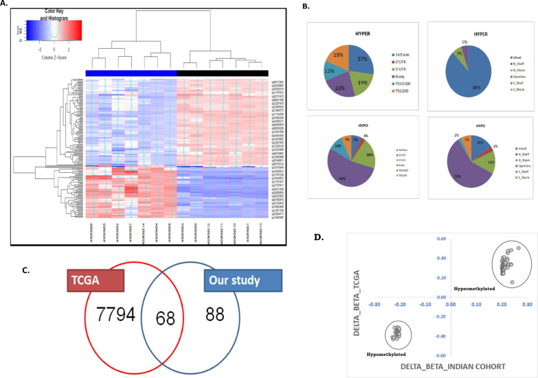
Fig. 1.
A Hierarchical clustering of the samples based on the top-ranked DMPs (n = 156): Illustrative heat map denoting the Hierarchical Clustering of the PDAC samples (n = 7) based on the top-ranked DMPs (n = 156). Blue to red denoted increase in beta value (hyper-methylation). The cancer samples were marked as Blue and the normal samples were marked as Black (B) Genomic annotations of DMPs-CpG islands (Islands, Shores (± 2 KB from the boundaries of the islands), Shelves (± 2 KB from the boundaries of the shores) and Open Sea) or the transcription start site (5ʹ UTR, Exon 1, Promoter, Body, Non-genic). Distribution of the hypo-methylated and hyper-methylated DMPs across different segments of the genome. C Venn diagram showing common and uncommon DMPs in the TCGA cohort and Indian cohort. Among the 7832 DMPs, 68 DMPs were common to our study finding. D Correlation plot between the delta beta values at these 68 DMPs in TCGA data (for 9 PAAD patients) and in Indian cohort (n = 7)

