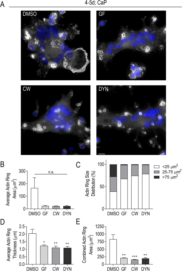
FIGURE 7:
Centrosome declustering drugs impact F-actin ring size and thickness. RAW-derived osteoclasts were lifted and replated on CaP prior to 24-h treatment with centrosome declustering agents: 40 μM GF, 80 μM CW069 (CW), and 175 μM DYN. (A) Representative immunofluorescent images of CaP-plated osteoclasts were stained for actin (gray) and DAPI (blue). Scale bars = 10 μm. (B, D, E) Average Actin Ring Area (μm2) (B), Average Actin Ring Thickness (μm) (D), and Combined Actin Ring Area (μm2) (E) was determined from three independent experiments (n = 40). Significance relative to DMSO was determined through a one-way ANOVA followed by Dunnett’s multiple comparison (*P < 0.05; **P < 0.01; ***P < 0.001). Each graph displays mean ± SEM. (C) Actin Ring Size Distribution (%) is presented as small (<25 μm2), midsize (25-75 μm2), and large rings (>75 μm2) from three independent experiments.

