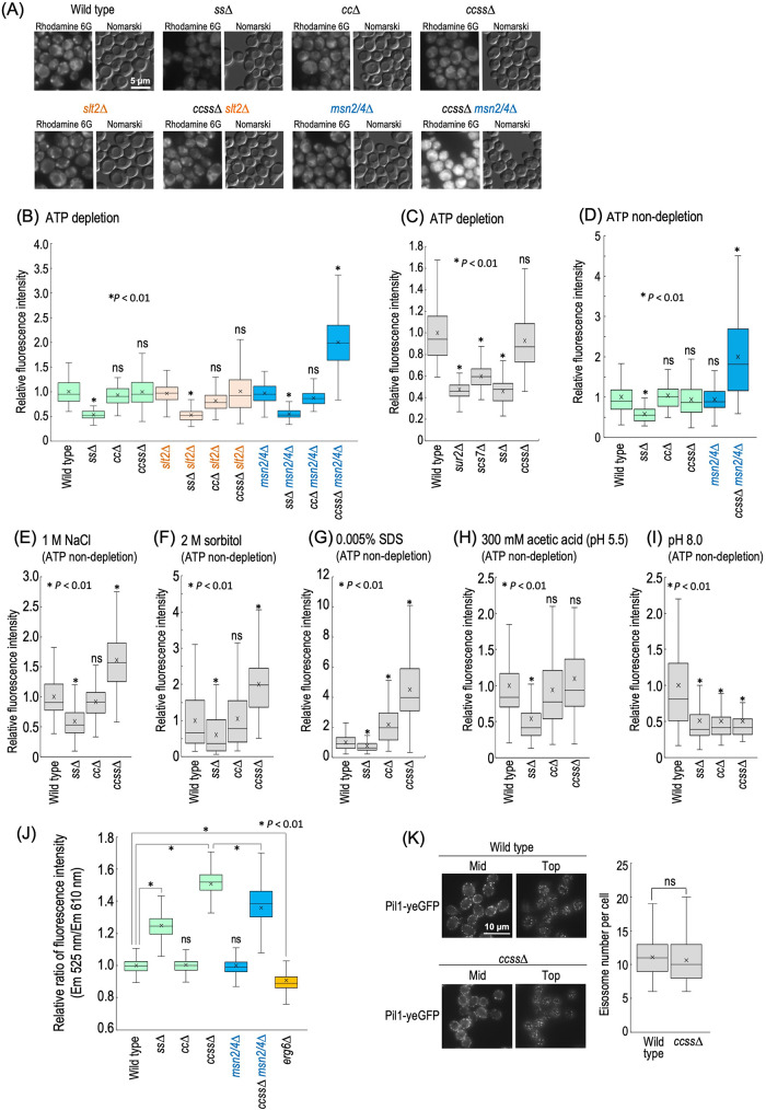
FIGURE 7:
Plasma membrane properties in ccss∆ cells. (A) Detection of rhodamine 6G incorporated into cells under ATP-depleted conditions. Cells were cultured overnight in YPD medium at 30°C, diluted (0.3 OD600 units/ml) in fresh YPD medium, and then incubated for 5 h at 30°C. Rhodamine 6G (10 µM) was added to cells (1 OD600 units/300 µl) under ATP-depleted conditions, followed by incubation for 30 min at 30°C. Cells were viewed under a fluorescence microscope. (B, C) Efficiency of incorporation of rhodamine 6G into cells under ATP-depleted conditions. The fluorescence intensity of individual cells is expressed as a boxplot. Data represent the value for 100 cells for individual strains. The average (marked as x) of fluorescence intensity in wild-type cells was taken as 1. ns: no significant difference. (D) Efficiency of incorporation of rhodamine 6G into cells under ATP-nondepleted conditions. (E–I) Efficiency of incorporation of rhodamine 6G under stress conditions. Cells (1 OD600 units/300 µl) were incubated with 10 µM rhodamine 6G in YPD medium containing 1 M NaCl (E), 2 M sorbitol (F), 0.005% SDS (G), or 300 mM acetic acid (adjusted to pH 5.5 by the addition of 50 mM MES and MOPS) (H) for 30 min. YPD medium buffered at pH 8.0 was prepared by the addition of 100 mM HEPES (I). (J) Evaluation of membrane lipid order by using di-4-ANEPPDHQ. Cells were cultured overnight in YPD medium at 30°C, diluted (0.3 OD600 units/ml) in fresh YPD medium, and then incubated for 5 h at 30°C. di-4-ANEPPDHQ (5 µM) was added to cells (1 OD600 units/100 µl), followed by incubation for 1 min at 30°C. The ratio of green (525 nm) and red (610 nm) fluorescences in individual cells, which was measured by a flow cytometer, is expressed as a boxplot. Data represent the value for 1000 cells for individual strains. The average (marked as x) of the fluorescence ratio in wild-type cells was taken as 1. (K) Distribution of eisosomes in wild-type and ccss∆ cells. Cells expressing Pil1-yeGFP were cultured overnight in YPD medium at 30°C, diluted (0.3 OD600 units/ml) in fresh YPD medium, and then incubated for 5 h at 30°C. Cells were observed under a fluorescence microscope. Eisosomes in top section of images of individual cells (n = 30) were counted by using Fiji (Schindelin et al., 2012; Sakata et al., 2022).

