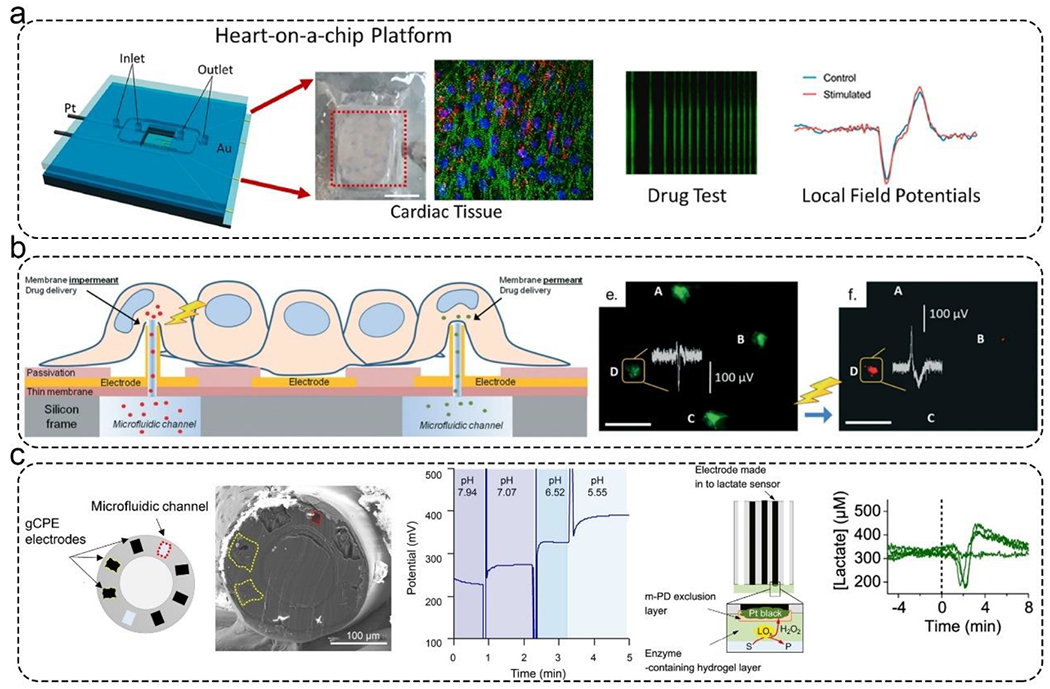
Figure 7.

Applications of microfluidic biosensors for monitoring MPSs. a). An integrated cardiac MPS with a platinum wire electrode for applying electrical stimulation, a gold MEAs for acquiring electrophysiological signals, and a microfluidic chamber for long-term culturing cells. This platform can test drug responses with local field potentials. Reproduced with permission from ref (138) Copyright 2021 Elsevier. b). An integrated cardiac MPS. The MEAs were decorated with 3D hollow nanostructures that could deliver calcein-AM and propidium iodide into cardiac cells. The MEAs could monitor this process. Reproduced with permission from ref (139) Copyright 2018 Royal Society of Chemistry. c). Potentiometric fiber electrodes were used to monitor pH and transient neurometabolic lactate in neural tissue. Left: Schematic illustration of the fiber-based biosensor. Middle: Real-time monitoring pH. Right: The detection principle of lactate sensor and the real-time monitoring lactate. Reproduced with permission from ref (142) Copyright 2021 American Chemical Society.
