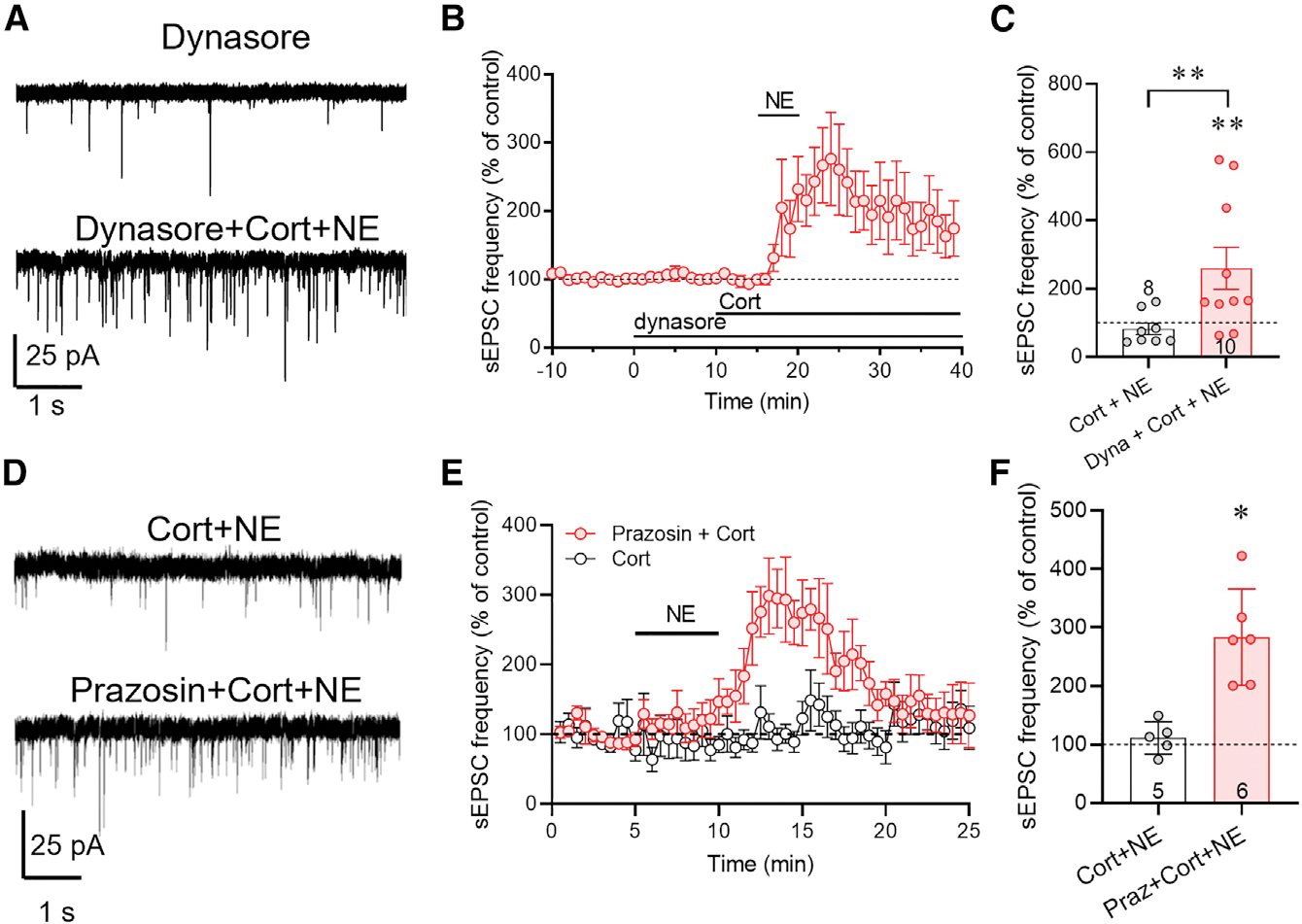
Figure 3. The corticosterone-induced desensitization of CRH neurons to NE excitation is dependent on ligand-mediated endocytosis.

(A) Representative recordings of sEPSCs recorded in CRH neurons at baseline in the dynamin inhibitor dynasore and after NE application following dynasore and corticosterone co-application (dynasore + Cort + NE).
(B) Time course of the change in normalized mean sEPSC frequency showing restoration of the NE-induced increase in sEPSC frequency by dynasore preapplication prior to co-application of Cort and NE.
(C) Summary of NE effect on normalized mean sEPSC frequency in corticosterone alone (Cort + NE) and in dynasore and corticosterone (Dyna + Cort + NE). Inhibiting endocytosis reversed the corticosterone blockade of the NE-induced increase in sEPSC frequency.
(D) Representative recordings of the sEPSC response to NE 3 h following preincubation of slices in corticosterone (Cort) and in the α1 adrenoreceptor antagonist prazosin and corticosterone (Prazosin + Cort).
(E) Time course of the normalized mean sEPSC frequency response to NE following corticosterone preapplication alone (Cort) and prazosin and corticosterone (Prazosin + Cort).
(F) Summary of the sEPSC frequency response to NE following pretreatment with corticosterone (Cort + NE) or with prazosin and corticosterone (Praz + Cort + NE). The corticosterone desensitization of the NE response was reversed by blocking α1 adrenoreceptors during corticosterone exposure. Data are represented as mean ± SEM; numerals in bar graphs represent numbers of recorded neurons. *p < 0.05, **p < 0.01.
See also Figure S2D.
