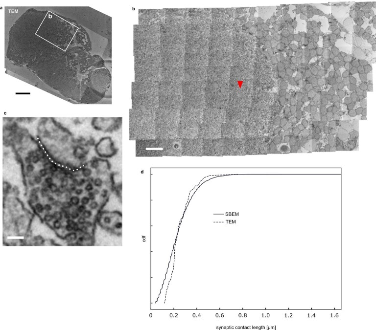
Extended Data Fig. 8. Comparison of synaptic contact lengths in TEM image of optic tectum.
a. Overview of 35 nm section of a 5 dpf larval zebrafish prepared identically to the sample used for SBEM. b. Close-up tile mosaic in optic tectum neuropil. Arrowhead points at synapse in (c). c. Example synapse. Dotted line illustrates synaptic contact length measurement. d. Distribution of contact lengths (n = 100, randomly sampled) in this TEM image (dotted line) compared to the distribution obtained from the optic tectum in the SBEM dataset (as in Extended Data Fig. 7b). Scale bars: a. 50 μm, b. 10 μm, c. 100 nm.

