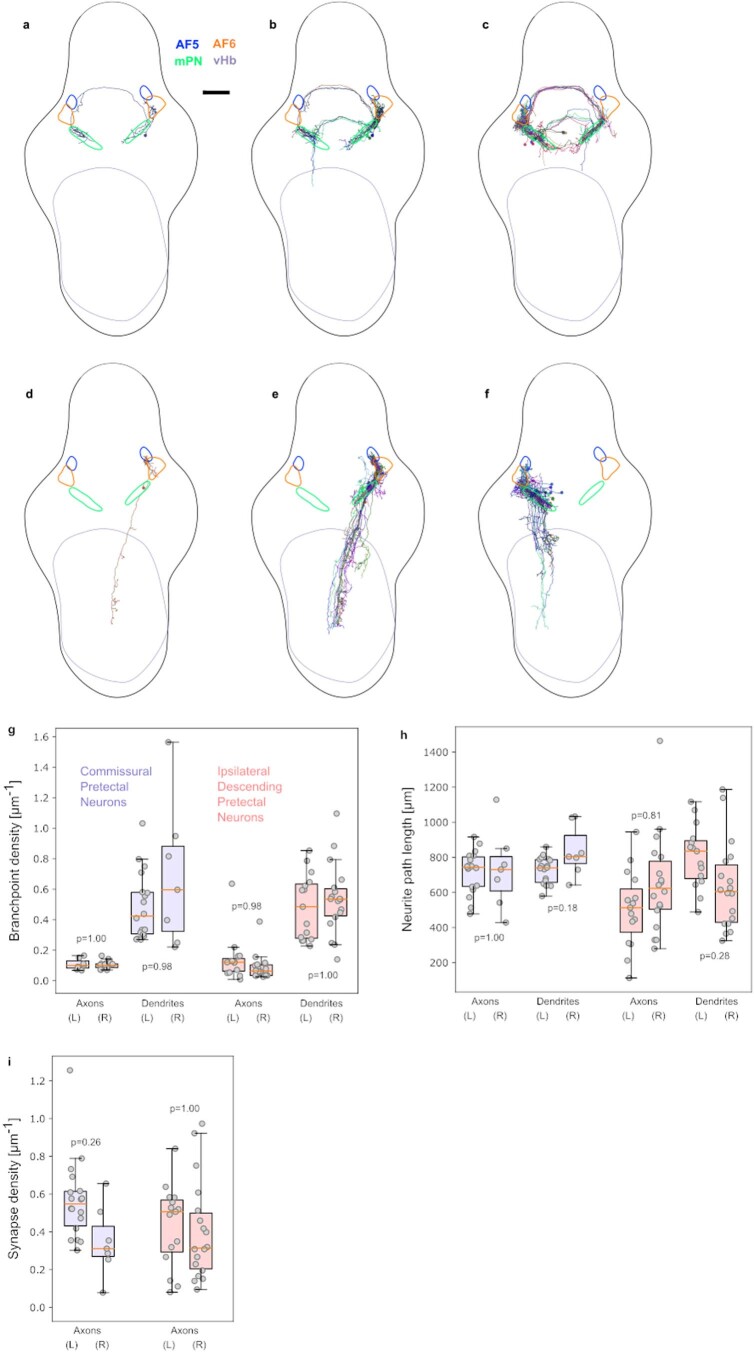
Extended Data Fig. 3. Bilaterally symmetric reconstructions of two morphologically defined pretectal cell types.
Individual example of a commissural pretectal interneuron (a) and all neurons of this type with somata on the right (b) and left (c) side of the brain. d-f. Like a-c, for ipsilateral descending pretectal projection neurons. g-i. Branchpoint densities (g), neurite path lengths (h) and axonal synapse densities (i) for commissural pretectal interneurons (blue, n = 18 and 7 cells on the left and right side, respectively) and ipsilateral descending pretectal projection neurons (red, n = 15 and 18 cells on the left and right side, respectively), plotted separately for axons and dendrites and for the two brain hemispheres. AF5, AF6: Retinal arborization fields 5 and 6, mPN: medial pretectal neuropil region, vHb: ventral hindbrain neuropil. AF annotations are based on LM atlas masks registered to the EM data. Box plot center lines represent medians, box limits upper and lower quartiles, whiskers 1.5x the interquartile range. Scale bar: 100 μm.

