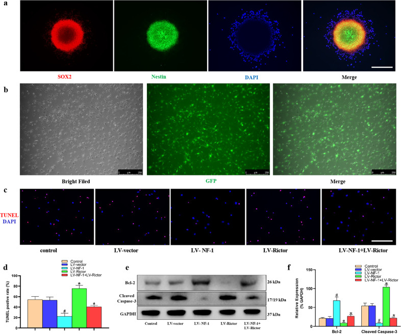
Fig. 1. Knockout of NF-1 improved the antiapoptotic ability of neural stem cells (NSCs) in vitro.
a NSCs gathered as neurospheres and abundantly expressed SOX2 and Nestin (scale bar = 100 µm). b After successful transfection by lentivirus, most of the NSCs expressed green fluorescent protein (GFP) (scale bar = 250 µm). c NSCs after inducing apoptosis were detected through terminal deoxynucleotidyl transferase-mediated dUTP nick-end labeling (TUNEL) (red: TUNEL-positive; blue: 4′-6-diamidino-2-phenylindole, DAPI) (scale bar = 100 µm). d Quantitative comparison of TUNEL-positive cells in different groups. e Western blotting of apoptosis-related proteins, including Bcl-2, cleaved caspase-3, and glyceraldehyde-3-phosphate dehydrogenase (GAPDH). f Quantitative comparison of the expression of Bcl-2 and cleaved caspase-3 in different groups (data are expressed relative to GAPDH). *P < 0.05 compared with the control and LV-vector groups; #P < 0.05 compared with the LV-NF-1+LV-Rictor group.

