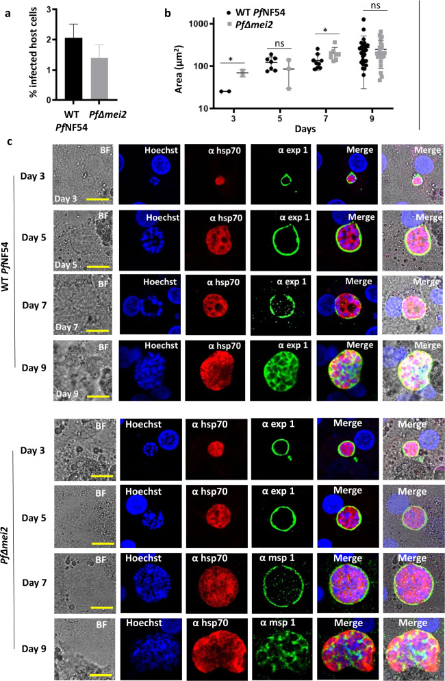
Fig. 2. PfΔmei2 liver-stage development in cultured human primary hepatocytes (BioIVT).
a Percentage of hepatocytes infected with WT PfNF54 and PfΔmei2 at day 3 post infection (p.i.) (p = 0.002; unpaired Mann–Whitney test). b Liver-stage size on day 3, 5, 7, and 9 p.i. (3-20 parasites measured in two wells). The average of the parasite’s cytoplasm at its greatest circumference using HSP70-positive area (μm2), s.d. and significances values are shown (unpaired Mann–Whitney test: *p < 0.05; ns: not significant). c Representative confocal microscopy images of liver stages on days 3, 5, 7, and 9 p.i. Upper panel WT PfNF54; lower panel PfΔmei2. Fixed hepatocytes were stained with the following antibodies: rabbit anti-PfHSP70 (α hsp70), mouse anti-PfEXP1 (α exp1), and anti-PfMSP1 (α msp1). Nuclei stained with Hoechst-33342. All pictures were recorded with standardized exposure/gain times; Alexa Fluor® 488 (green) 0.7 s; anti-IgG Alexa Fluor® 594 (red) 0.6 s; Hoechst (blue) 0.136 s; bright field (BF) 0.62 s (1× gain). Scale bar, 10 μm. Error bars represent standard deviation.

