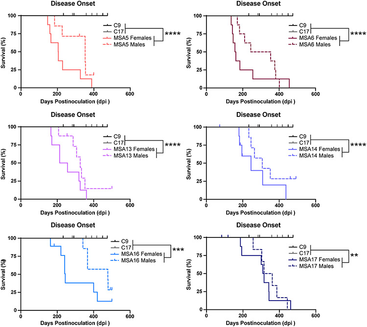
Fig. 1.
MSA patient samples transmit neurological disease to TgM20+/− mice. Eight-week-old TgM20+/− mice were inoculated with either 30 μL of two control patient samples (C9 and C17) or six MSA patient samples (MSA5, MSA6, MSA13, MSA14, MSA16, and MSA17). Kaplan–Meier plots show disease onset in female (solid line) and male (dotted line) TgM20+/− mice. Control-inoculated mice did not develop disease by 475 days postinoculation (dpi), but mice inoculated with all six MSA samples developed progressive neurological signs. Incubation times shown in Table S2, online resource (** = P < 0.01; *** = P < 0.001; **** = P < 0.0001). Female mice inoculated with MSA patient samples developed neurological disease ~ 63 days earlier than male mice (P = 0.0005)

