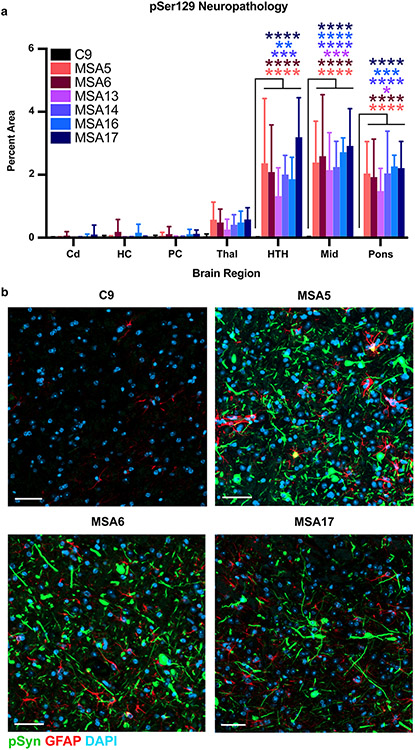
Fig. 2.
MSA patient samples induce phosphorylated α-synuclein neuropathology in terminal TgM20+/− mice. Eight-week-old TgM20+/− mice were inoculated with 30 μL of C9 control sample or six MSA patient samples (MSA5, MSA6, MSA13, MSA14, MSA16, and MSA17). a Quantification of stained brain slices showed no phosphorylated α-synuclein inclusions were present in the caudate (Cd), hippocampus (HC), piriform cortex and amygdala (PC), thalamus (Thal), hypothalamus (HTH), midbrain (Mid), or pons of C9-inoculated mice. However, the MSA patient samples induced neuropathological lesions in the HTH, Mid, and pons (* = P < 0.05; ** = P < 0.01; *** = P < 0.001; **** = P < 0.0001). b Representative images of the Mid from TgM20+/− mice inoculated with C9, MSA5, MSA6, or MSA17 brain homogenate. Phosphorylated α-synuclein in green, GFAP in red, and DAPI in blue. Scale bar: 50 μm

