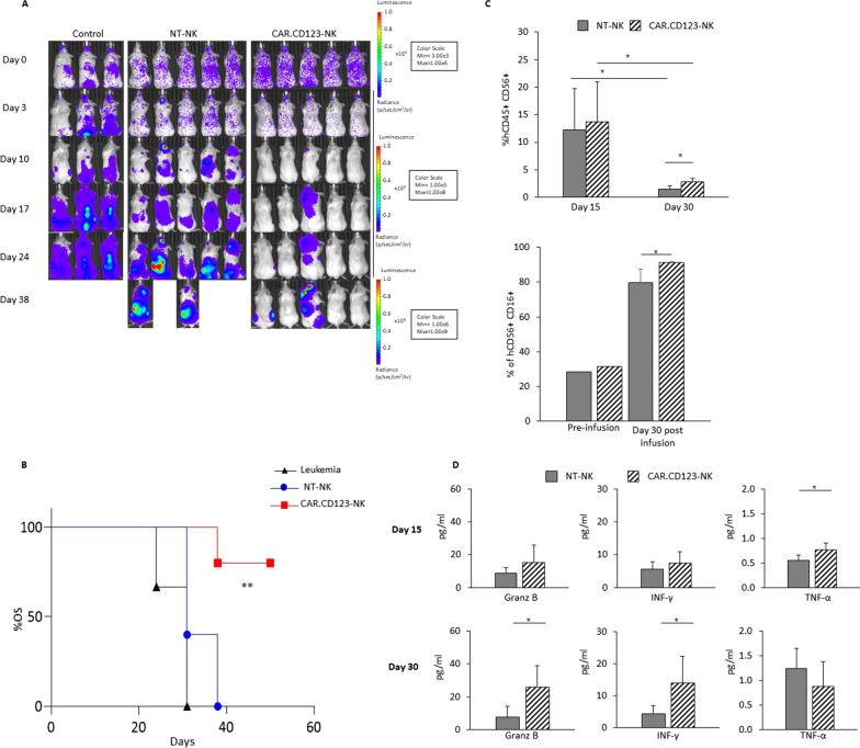
Fig. 6.
CAR.CD123-NK cells maintain efficacy and safety profile in an AML xenograft animal model based on hIL15-NOG mice. A hIL15-NOG mice were systemically engrafted with THP-1-FF-Luc.GFP cells and, at the time of leukaemia engraftment, treated or not with NT-NK and CAR.CD123-NK cells. Time course of in vivo bioluminescence imaging of the treated NOG mice. B Fifty-day probability of OS for NSG mice systemically infused with THP-1-FF-Luc.GFP cells after adoptive transfer of NT-NK (blue bars with circles) or CAR.123-NK cells (red bars with squares). The untreated control is represented by a black line with triangles. C Flow cytometry analysis of human NK cells in PB of mice (n = 5 mice receiving NK-NT; n = 5 mice receiving CAR.CD123-NK) performed at Day 15 and Day 30 after effector cell infusion. Plots show the percentage of hCD45+ hCD56+ hCD3− cells gated on the total cells present in PB of the mice (left panel) and as the percentage of hCD45+ hCD16+ cells of the total hCD45+ hCD56+ CD3.− cells present in PB of the mice (right panel). Data are shown as average ± SD. *p < 0.05. D Cytokine concentration was measured in the plasma of mice bearing THP-1-FF-Luc.GFP cells and treated with NT-NK (grey bars) and CAR.CD123-NK (black line bars) cells at Day 15 and at Day 30 after effector cell infusion. Granz B, IFN-γ and TNF-α were measured by ELISA assay, and data are shown as average ± SD. *p < 0.05, **p < 0.01

