FIG. 6.
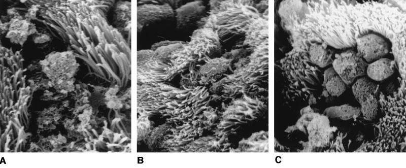
Scanning electron micrographs of human fallopian tubes infected for 24 h with gly1 mutant N. gonorrhoeae strains expressing gly1 in trans. Tissues were infected and examined as in Fig. 6. (A) Tissue infected with the gly1 mutant strain 120 (magnification, ×6,636). (B) Tissue infected with the complemented mutant, 120(pGly18.2.2) (magnification, ×3,634). (C) Tissue infected with MS11A(pGly18.2.2) (magnification, ×3,634).

