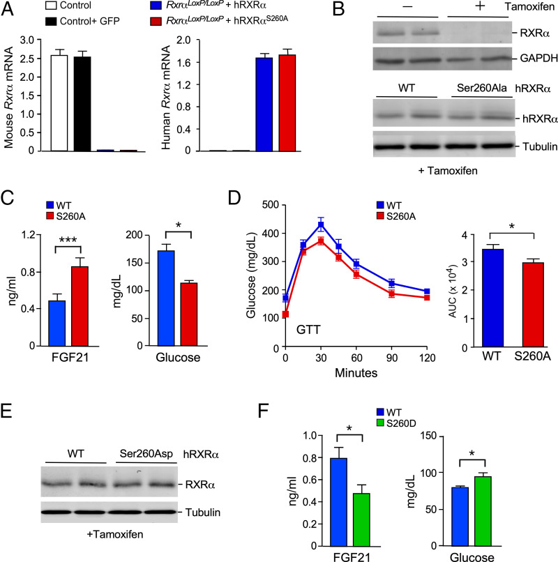
Fig. 3.
Hepatic hRXRα phosphorylation on Ser260 increases FGF21 signaling. (A) Control SA-CreERT2 mice and SA-CreERT2 RxraloxP/loxP mice (age 7 wk) were transduced with AAV8 viruses expressing green fluorescent protein (GFP), hRXRα, or Ser260Ala-hRXRα. The mice were treated without or with tamoxifen at age 8 wk, fed a HFD starting at age 10 wk, and euthanized at age 18 wk. Reverse transcriptase quantitative PCR (RT-qPCR) assays of murine liver to detected mouse and human Rxra mRNA are presented (mean ± SEM; n = 8∼10). (B) Lysates of murine liver were examined by immunoblot analysis by probing with antibodies to RXRα, glyceraldehyde-3-phosphate dehydrogenase (GAPDH), and α-tubulin. (C) Fasting blood FGF21 and glucose concentration in HFD-fed mice expressing hRXRα or Ser260Ala hRXRα in hepatocytes was measured (mean ± SEM; *P < 0.05, ***P < 0.001; n = 9∼10). (D) GTTs on HFD-fed mice expressing hRXRα or Ser260Ala hRXRα in hepatocytes were performed and the area under the curve (AUC) was measured (mean ± SEM; *P < 0.05; n = 7∼10). (E) CD-fed mice expressing WT hRXRα or Ser260Asp hRXRα were euthanized at age 18 wk. Immunoblot analysis of liver extracts was performed by probing with antibodies to RXRα and α-tubulin. (F) Fasting blood FGF21 and glucose concentration in CD-fed mice expressing hRXRα or Ser260Asp hRXRα in hepatocytes was measured (mean ± SEM; *P < 0.05; n = 9).

