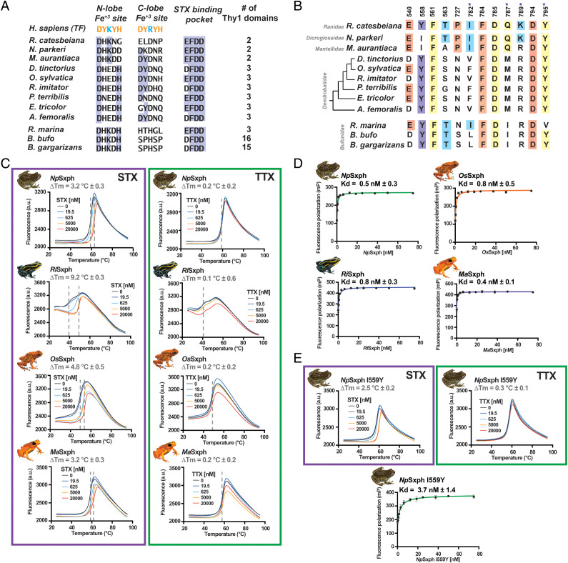
Fig. 5.
Sxph family member properties. (A) Comparison of the human transferrin (TF) Fe3+ ligand positions (UniProtKB: P02787), RcSxph STX binding motif residues (10), and number of Thy1 domains for Sxphs from R. castesbeiana (PDB ID:6O0D) (10), N. parkeri (NCBI: XP_018410833.1) (10), M. aurantiaca, D. tinctorius, O. sylvatica, R. imitator, P. terribilis, E. tricolor, A. femoralis, R. marinus, B. bufo (NCBI:XM_040427746.1), and B. garagarizans (NCBI:XP_044148290.1). TF Fe3+ (orange) and carbonate (blue) ligands are indicated. Blue highlights indicate residue conservation. (B) Comparison of STX binding pocket for the indicated Sxphs. Numbers denote RcSxph positions. Colors indicate the alanine scan classes as in Fig. 3B. Conserved residues are highlighted. Asterix indicates second-shell sites. (C) Exemplar TF curves for NpSxph, RiSxph, OsSxph, and MaSxph in the presence of the indicated concentrations of STX (purple box) or TTX (green box). ΔTm values are indicated. (D) Exemplar FP binding curves and Kds for NpSxph (green), RiSxph (blue), OsSxph (orange), and MaSxph (purple). (E) Exemplar NpSxph I559Y TF curves in the presence of the indicated concentrations of STX (purple box) or TTX (green box) and FP binding (green). ΔTm and Kd values are indicated. Error bars are SEM.

