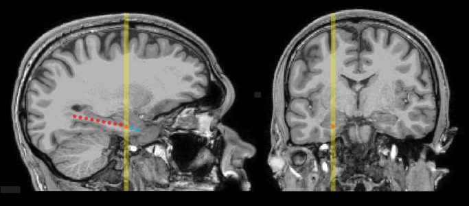
Fig. 4.
MRI scans showing locations of medial-temporal electrodes in a representative patient (P1) in sagittal and coronal planes. Depth probes were implanted through a small hole in the skull and targeted for the hippocampus, guided by anatomical information from pre-surgical MRI. Contacts were cylindrical, 1.25 mm in diameter and 2.5 mm long, situated 5 mm apart along each probe (measured center-to-center). The image on the left shows a sagittal section that includes the estimated location of the contact selected for primary iEEG analysis as an orange circle. Locations of other contacts within the section are shown as red circles. Locations of contacts in adjacent sagittal sections were projected onto approximate locations in the section and shown as open blue circles. The yellow vertical line on each image indicates the location of the selected contact and corresponds to the approximate level of the section opposite.

