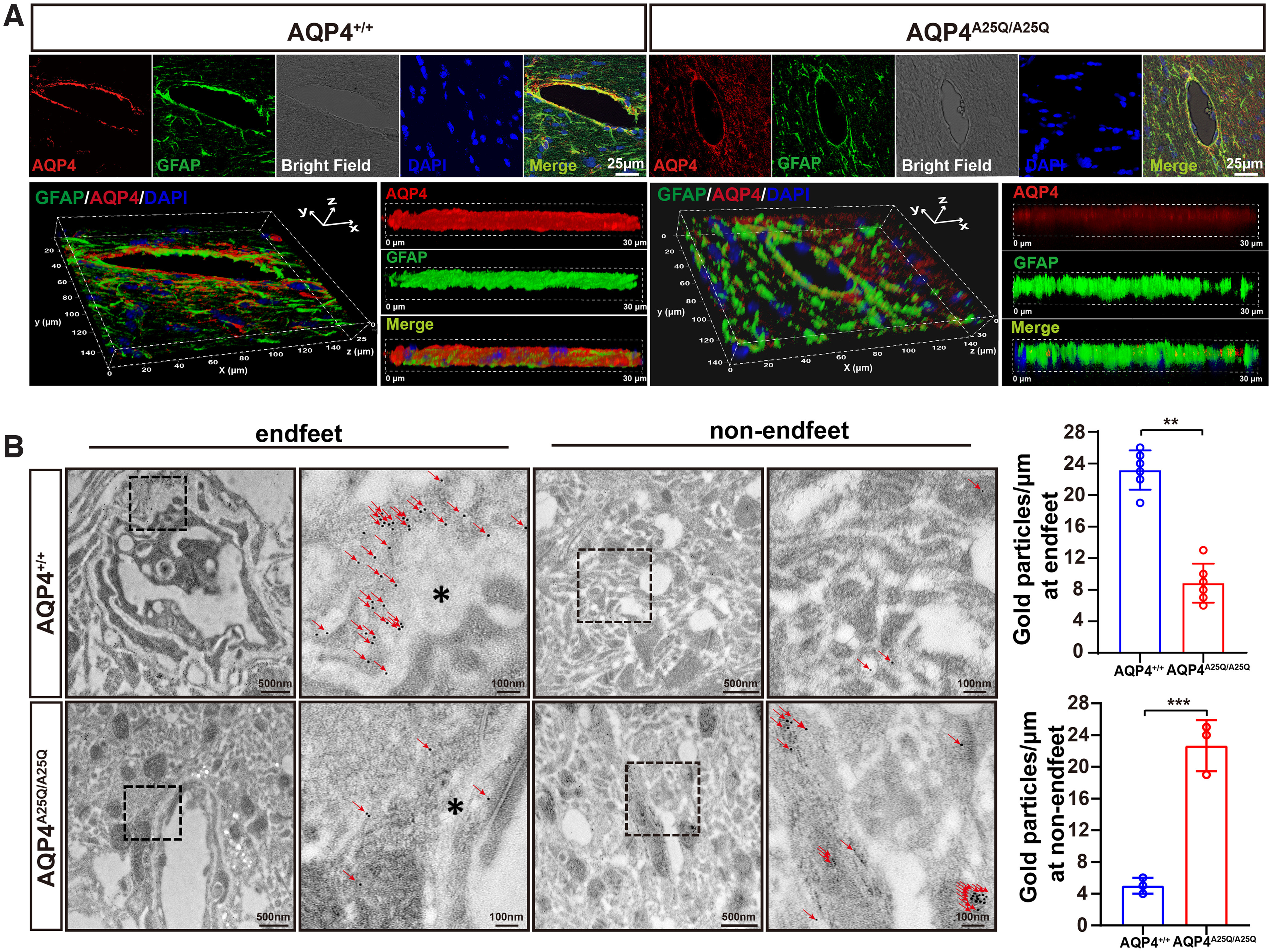
Figure 4.

AQP4-A25Q mutation decreases polarized AQP4 expression in glial endfeet. A, Distribution of AQP4 in astrocytes of AQP4+/+ and AQP4A25Q/A25Q mice. Coimmunostaining with anti-AQP4 (red) and anti-GFAP (green) antibodies with DAPI staining for cell nucleus was performed. Bright field represents the microvasculature. Scale bar: 25 μm. 3D reconstruction of a confocal z stack of AQP4+/+ and AQP4A25Q/A25Q mouse brain slices. Scale bar: 30 μm. B, Left, Representative locations of AQP4 in AQP4+/+ and AQP4A25Q/A25Q mice indicated by IEM staining. In AQP4+/+ mice, gold particles (red arrowheads) were concentrated in astrocyte endfeet (asterisks), whereas in AQP4A25Q/A25Q mice, AQP4 labeling was scattered. Scale bars: 100 nm, 500 nm. Right, Quantitative analysis of AQP4 immunogold particles labeling in AQP4+/+ and AQP4A25Q/A25Q mice. **p<0.01; ***p<0.001; Mann–Whitney test. n = 6 mice for gold particles at endfeet. n = 3 mice for gold particles at non-endfeet region. Bar graphs represent the mean ± SD. Antibodies used are provided in Extended Data Table 2-1.
