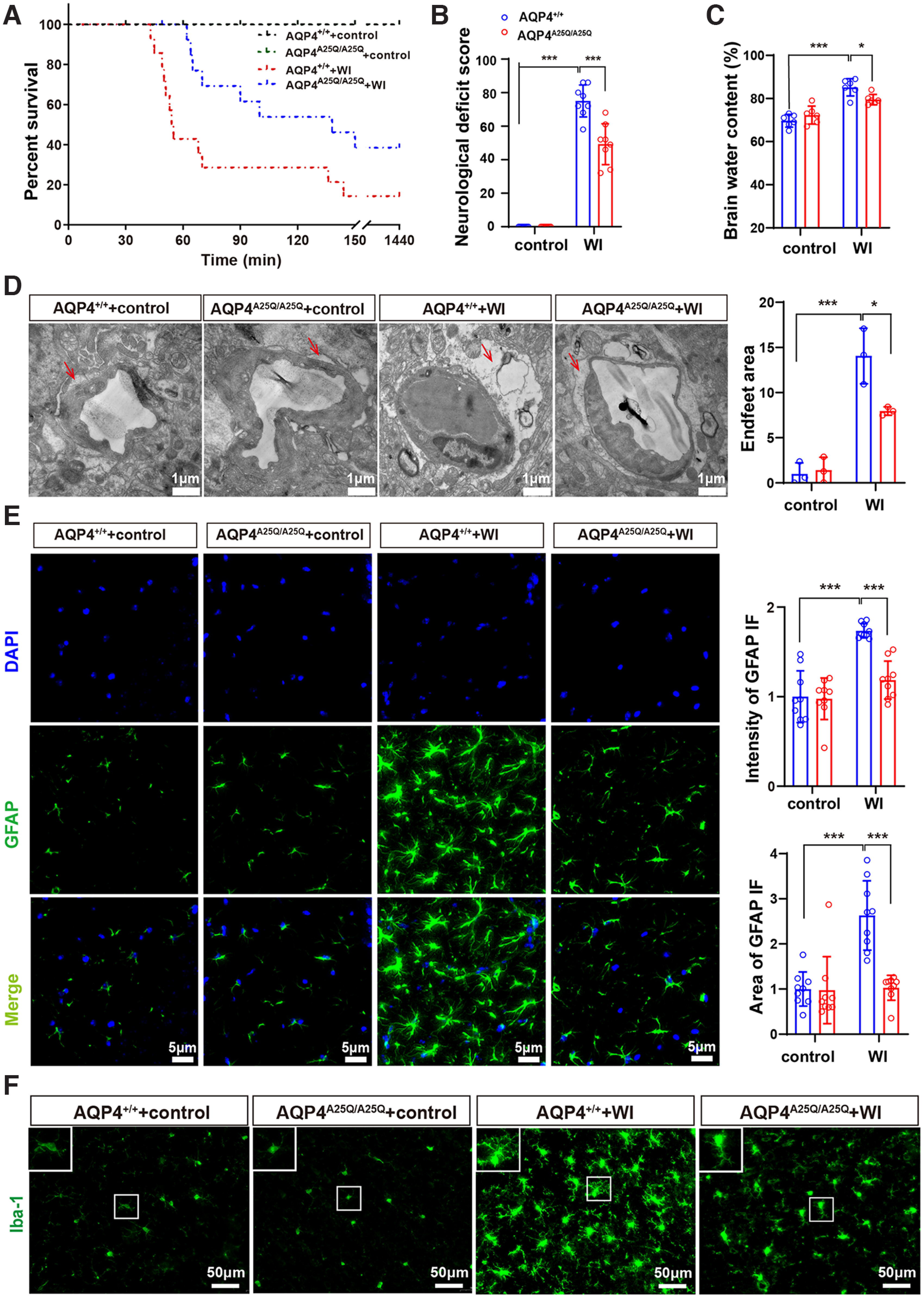
Figure 5.

AQP4A25Q/A25Q mice exhibited improved brain edema, neurologic outcomes, and survival after water intoxication (WI). A, Cumulative survival of AQP4+/+ and AQP4A25Q/A25Q mice with and without WI; n = 14 mice. B, Neurologic scores of AQP4+/+ and AQP4A25Q/A25Q mice with and without water intoxication; n = 8 mice. C, Brain water contents of AQP4+/+ and AQP4A25Q/A25Q mice with and without water intoxication; n = 6 mice. D, Left, Transmission electron micrograph represents an edematous cerebral cortex at 30 min after water intoxication, the red arrow indicates astrocyte endfeet process. Scale bar, 1 μm. Right, Quantification of the pericapillary astrocyte endfeet process area was performed with 3 fields/mouse; n = 3 mice for each group. E, Left, Representative photographs of GFAP staining in the astrocytes of AQP4+/+ and AQP4A25Q/A25Q mice with and without water intoxication. Scale bar, 5 μm. Right, The intensity and area of GFAP in the brain were divided by the number of cells counterstained with DAPI; n = 9 mice for each group. Total number of GFAP-positive cells in control and mutant mice with water intoxication is provided in Extended Data Figure 5-1. F, Immunofluorescence staining of Iba-1 in AQP4+/+ and AQP4A25Q/A25Q mice with and without water intoxication. Scale bar, 50 μm. Expression of AQP4 in a different group of mice is provided in Extended Data Figure 5-2. A, Log-rank (Mantel–Cox) test was used. B-E, Data are mean ± SD. *p < 0.05; ***p<0.001; two-way ANOVA with Tukey's post hoc test.
