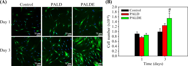
FIG. 3.
Schwann cell proliferation assay. (a) Confocal microscopic images of SCs stained with DAPI (4′,6-diamidino-2-phenylindole) (blue) and S100 (green) to observe SC proliferation on days 1 and 3. (b) Quantification of SC proliferation in control, PALD, and PALDE groups. Data are presented as mean ± standard deviation. *p < 0.05 compared with the control group. #p < 0.05 compared with the PALD group. PALD, pluronic–alginate–lysine–dextran; PALDE, pluronic–alginate–lysine–dextran loaded with extracellular vesicles.

