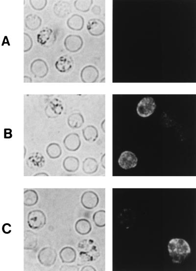
FIG. 4.
Immunofluorescence staining of fixed smears of erythrocytes infected with P. yoelii. Thin blood smears were incubated with preimmune rabbit serum (A) or rabbit antiserum raised against recombinant pAg-1N (B and C), followed by a fluorescein isothiocyanate-conjugated secondary antibody. The same fields of cells were examined by phase-contrast (left panels) and fluorescence (right panels) microscopy.

