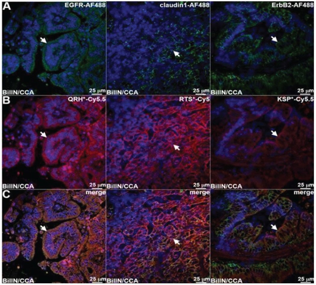
Figure 5: Co-localization of antibody and peptide binding to biliary neoplasia.

A) Fluorescence images collected with confocal microscopy show AF488-labeled antibodies (green) specific for EGFR, claudin-1, and ErbB2 binding to the cell surface (arrows) of neoplastic biliary epithelial cells. B) Peptides labeled with either Cy5.5 or Cy5 (red) also show cell surface binding (arrows) to adjacent sections. C) Merged images show co-localization of binding (arrows) with a Pearson’s correlation coefficient of ρ = 0.64, 0.51 and 0.62 for EGFR, claudin-1, and ErbB2 respectively.
