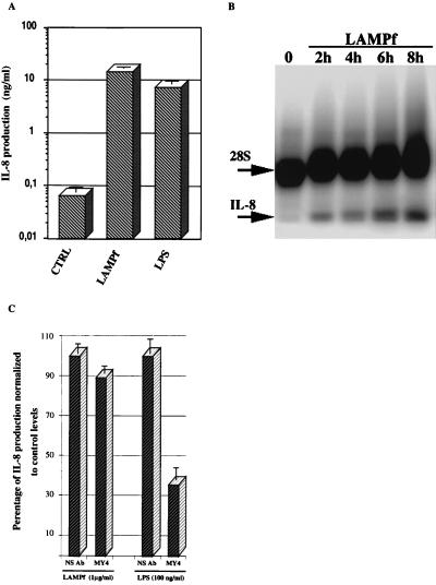
FIG. 1.
IL-8 production by human THP-1 cells in response to LAMPf and LPS. (A) THP-1 cells (106/ml) were not stimulated (control [CTRL]) or stimulated with either LPS (1 μg/ml) or LAMPf (1 μg/ml). IL-8 production was measured by ELISA 18 h after stimulation. Data presented are means ± SEM of three distinct assays. (B) Multi-NPA using oligonucleotide probes for IL-8 and 28S rRNA (for standardization). RNAs were prepared from untreated THP-1 cells (○) and THP-1 cells induced with LAMPf (1 μg/ml) for the indicated time and subjected to multi-NPA analysis. Multi-NPA gels were exposed to a PhosphorImager screen for 2 to 4 h; the gel shown is representative of three experiments with similar results. IL-8 and 28S rRNA oligonucleotide probes protect 24 and 35 bases of the IL-8 and 28S rRNA transcripts, respectively. The IL-8 signal in each lane was quantitatively assessed and normalized to the 28S rRNA signal. (C) Effect of anti-CD14 antibody treatments on LAMPf-induced IL-8 secretion. Human THP-1 cells were incubated with anti-human CD14 monoclonal antibody MY4 at 5 μg/ml for 1 h prior to stimulation with either LAMPf (1 μg/ml) or LPS (100 ng/ml). An irrelevant antibody (NS) was used as a control. IL-8 secretion was determined after 18 h of culture and expressed as percentage of secretion normalized to stimulated cells that received no antibody treatment. Mean values of two different experiments are shown.

