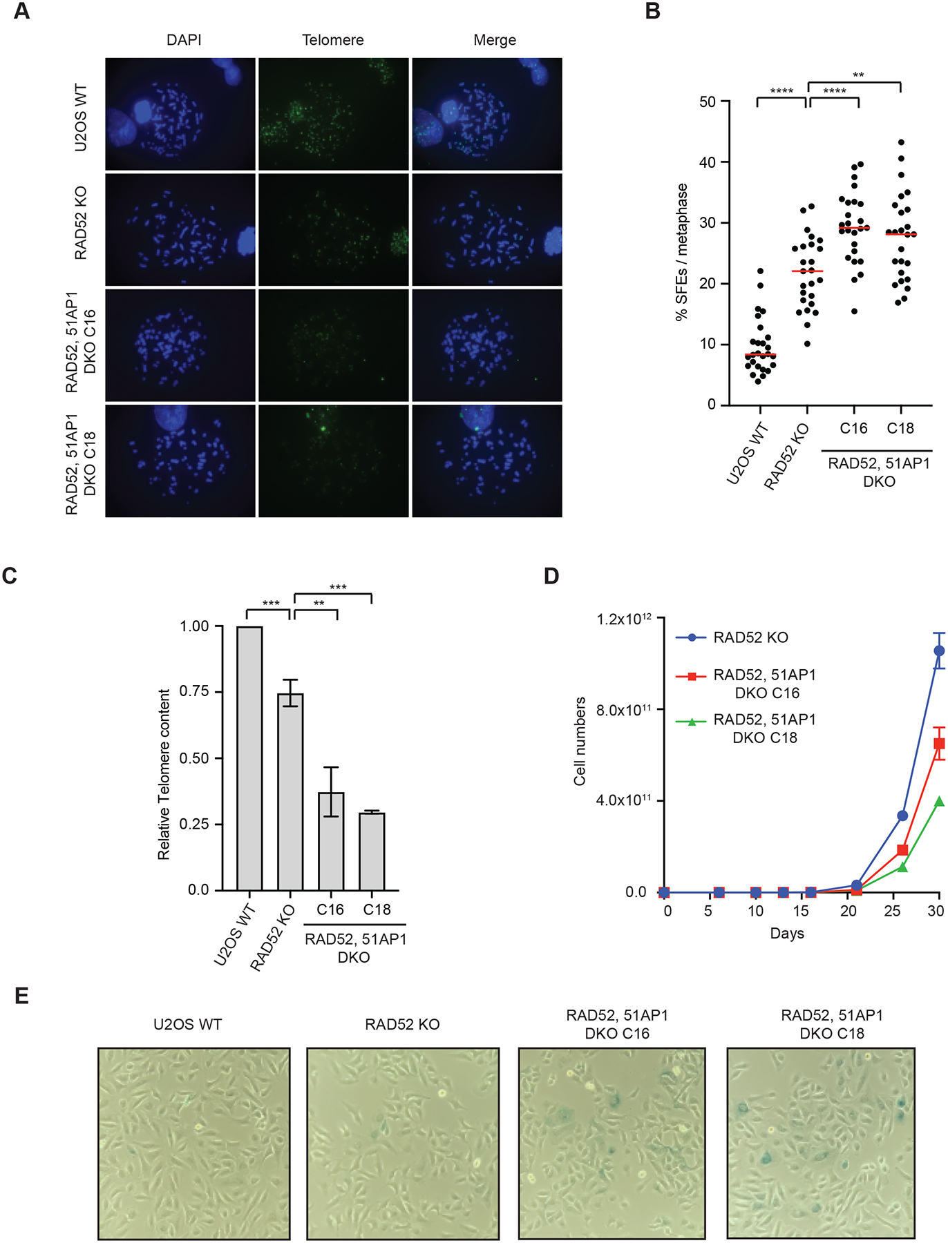
Figure 3. RAD51AP1 promotes telomere maintenance and proliferation in RAD52 KO cells.

(A) U2OS WT, RAD52 KO, and RAD52, 51AP1 DKO (clones C16 and C18) cells were synchronized in metaphase and telomeric FISH was performed using TelC-488 probe. (B) Signal free ends (SFEs) were quantified from (A). >25 metaphases were quantified from each cell line. Red lines: mean values. ****: P value <0.0001, **: P value 0.003. (C) Telomeres DNA content was quantified in U2OS WT, RAD52 KO, and RAD52, 51AP1 DKO (clones C16 and C18) cells after 1 month of passages. Error bars: SD, n=3 (experimental triplicates); ***: P value 0.001; **: P value 0.003 (D) Cell growth analysis of U2OS WT, RAD52 KO, and RAD52, 51AP1 DKO (clones C16 and C18) cells. (E) Representative images of β -galactosidase staining of U2OS WT, RAD52 KO, and RAD52, 51AP1 DKO (clones C16 and C18) cell populations. All cell lines were passaged in parallel for ~1 month.
