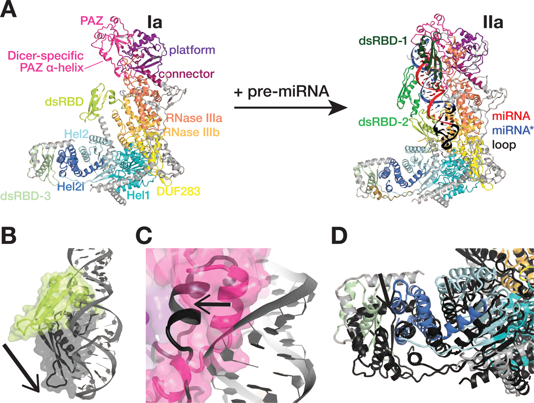
Figure 2. Dcr-1•Loqs-PB conformational equilibrium strongly favors closed states.
(A) Overall views of the RNA-free Dcr-1•Loqs-PB (structure Ia) and pre-miRNA-bound Dcr-1•Loqs-PB in presence of Ca2+ (structure IIa).
(B–D) Close-up views showing differences in the positions of the Dcr-1 dsRBD (B), the Dicer-specific α-helix of the PAZ domain (C), and the Dcr-1 Hel2i and the Loqs-PB dsRBD-3 domains (D) between the RNA-free structure Ia (colored) and the RNA-bound structure IIa (protein domains in dark gray and pre-miRNA in gray). Structural alignments were performed by superposition of Dcr-1•Loqs-PB complexes. Black arrows highlight conformational changes upon pre-miRNA binding.
See also Figure S5.

