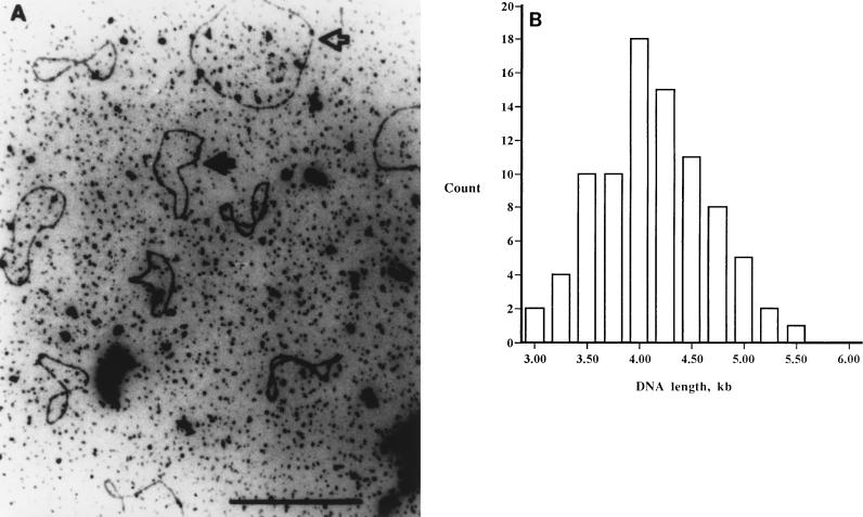
FIG. 1.
(A) Electron micrograph demonstrating that the pyocin DNA is a single-stranded closed circle. The open arrow designates M13, and the solid arrow identifies the pyocin ssDNA. (B) Distribution of the sizes of 86 pyocin ssDNA circles. M13 ssDNA was included in the sample, and measurement of 30 M13 circles served as the standard for estimating the size of the pyocin ssDNA.

