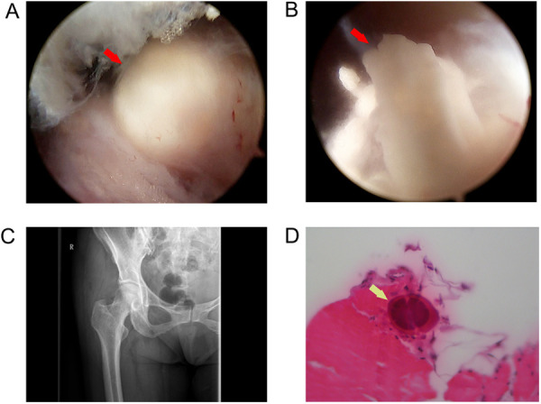
Figure 3.

(A) Endoscopic view of a calcified deposit, (B) Endoscopic view showing a soft white toothpaste-like material in the deposit, (C) postoperative plain radiograph view showing complete removal of the calcific deposit, and (D) hydroxyapatite crystals in the specimen confirmed by a pathologic examination (marked with arrows).
