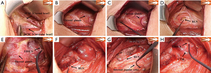
Figure 1.
Step-by-step description of SMIA. (A) Dissect between the branches of the SCM (between the sternal and clavicular heads) upward to the level of the larynx; (B) separate and exposure the affected thyroid gland at the deep surface of the strap muscle to reach the midline; (C) vagal signals monitored and recorded as V1; (D) “cross” method used to locate and protect the bilateral cervical RLNs, and the signal value R1 of nerve monitoring is recorded; (E) exposure and monitoring of the EBSLN; (F) after thyroid gland resection, the nearest end of the RLN is tested and recorded as EMG signal R2; (G) after complete hemostasis, the EMG signal of the vagus nerve V2 is obtained and recorded; (H) after thyroid gland resection, monitoring of the EBSLN. SMIA, sternocleidomastoid intermuscular approach; SCM, sternocleidomastoid muscle; RLN, recurrent laryngeal nerve; EBSLN, external branch superior laryngeal nerve; CTM, cricothyroid muscle; EMG, electromyographic.

