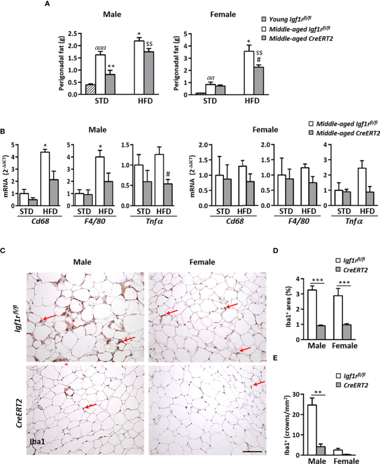
Figure 8.
Effects of IGF1R deficiency on perigonadal adipose tissue content and inflammation in middle-aged mice on HFD diet. (A) Effects of IGF1R depletion on perigonadal fat weight in male and female mice fed with either STD or HFD compared to young control (Igf1rfl/fl ) male and females on STD (n= 4-10 mice per group). (B) mRNA levels of inflammatory markers Cd68, F4/80 and Tnfα mRNA levels in midlle-aged mice fed either with STD or HFD. Gene expression is expressed as fold changes (2-ΔΔCt) compared to its Igf1rfl/fl corresponding control group. Data are expressed as mean ± SEM of at least 4 animals per group. (C) Representative immunohistochemical stainings of perigonadal fat tissue for the specific macrophage marker Iba1 (red arrows) in middle-aged mice fed with HFD. Crown-like structures, composed of macrophages surrounding adipocytes, are indicated with red asterisks. Scale bar: 100 µm. (D) Graphical representation of Iba+ stained area quantification (percentage) shown in (C). (E) Quantification of crown-like Iba1+ structures (density). Data in (D, E) are expressed as mean ± SEM of 2-10 animals per group (data obtained per animal analyzed in duplicated sections). aa p < 0.01 and aaap < 0.001 vs. young Igf1rfl/fl mice; *p < 0.05 and **p < 0.01 vs. middle-aged Igf1rfl/fl STD animals; #p < 0.05 vs. middle-aged Igf1rfl/fl HFD middle-aged mice; and $$ p < 0.01 vs. middle-aged CreERT2 STD animals in (A, B) **p < 0.01 and ***p < 0.001 vs. corresponding Igf1rfl/fl mice, in (D, E).

