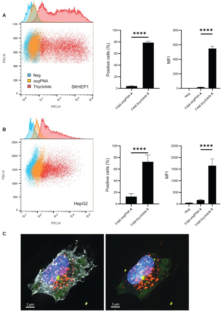
Figure 3.
Thyclotide 5 is successfully taken up by cells and does not colocalize with endosomes 16 hours after treatment. (A) FACS of SKHEP1 cells either non-treated (blue) or treated with 5 μM of either FAM-aegPNA 4 (orange) or FAM-thyclotide 5 (red). Positive cells were determined by having a fluorescence higher than the non-treated cells. MFI refers to the mean fluorescence intensity. (B) FACS of HepG2 cells either non-treated (blue) or treated with 5 μM of FAM-aegPNA 4 (orange) or FAM-thyclotide 5 (red). Positive cells were determined by having a fluorescence higher than the non-treated cells. MFI refers to the mean fluorescence intensity. (C) 3D volume reconstruction of 0.15 μm z-steps super-resolution microscopy imaging of SKHEP1 cells treated with FAM-thyclotide 5. Cells were stained with Membrite Fix 640/660 cell membrane marker (white), CellLight BacMam 2.0 early endosomes-RFP marker (orange) and Hoechst 33342 nucleus staining solution (blue). The membrane marker channel was removed from the right picture for a better visualization of the cytoplasmic diffusion of FAM-thyclotide 5 (green).

