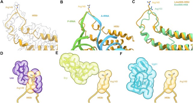
Figure 3.
Interaction of the NTD subdomain II of HflXr at the PTC. (A) Arg149 of NTD2-loop of HflXr (orange) in extracted cryo-EM density. (B) HflXr loop (orange) superimposed with A- (blue) and P-site tRNAs (green)(73). (C) The loop of HflXr (orange) extends deeper into the PTC than E. coli HflX (17). Alignment in (B) and (C) are based on 23S rRNA. D-F, Binding position of HflXr NTD2-loop (orange) with Arg149 shown as sphere, superimposed with the binding site of (D) Lincomycin (Lnc, purple, PDB ID 5HKV)(57), (E) erythromycin (Ery, yellow, PDB ID 4V7U) (59) and (F) virginiamycin S1 (VgS1, blue, PDB ID 1YIT)(60). Predicted steric clashes are indicated by red lines at the overlap of spheres.

