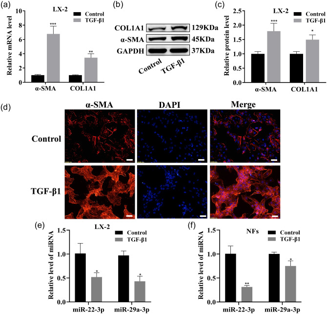
Figure 2.
miR-22-3p and miR-29a-3p expressions in activated LX-2, activated NFs. (a to c) qRT–PCR and Western blotting were used to detect COL1A1 and α-SMA mRNA and protein expression in LX-2. (d) IF images of activated LX-2 stained for α-SMA (red, ×200 magnification). Scale bars = 50 μm. (e and f) These two miRNAs were tested by qRT–PCR in LX-2 and NFs. (A color version of this figure is available in the online journal.)

