. 2022 Nov 7;12(49):31747–31756. doi: 10.1039/d2ra02593k

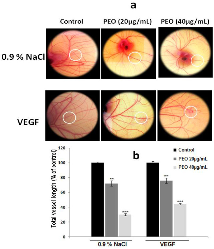
Fig. 7. PEO inhibits in vivo angiogenesis. (a) The CAM models were prepared using 8 days-old chick embryos. White circles represent location of applied disks. After incubation for 48 h with or without PEO extract, CAMs were photographed with a digital camera. Each group contained 3 CAMs and the experiment was repeated three times. (b) The quantitative measurement of total vessel length was performed on 50% of the total CAM surface treated in the absence or in the presence of PEO extract. For total vessel length, results are expressed as means ± SD of three experiments performed in triplicate and difference between treated and untreated (control) cells was considered statistically significant at **P < 0.01; ***P < 0.001.

Fig. 7