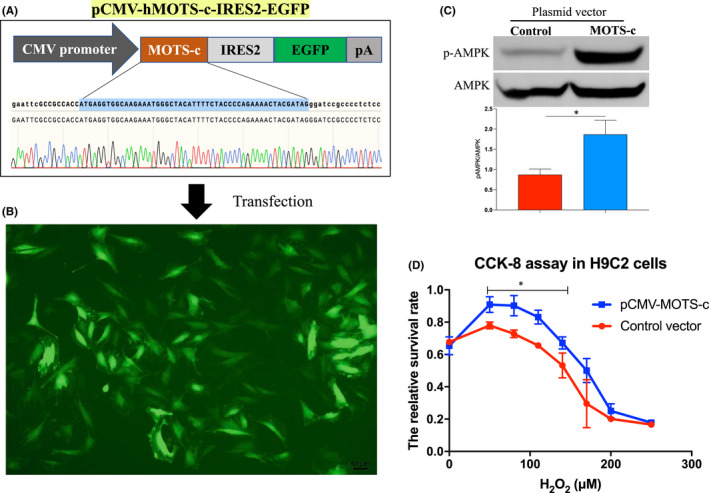
FIGURE 4.

MOTS‐c peptide protected cell apoptosis in H9C2 cells in response to oxidative injury. (A) Schematic images of human MOTS‐c (hMOTS‐c) expressing plasmid vector (pCMV‐hMOST‐c‐IRES2‐EGFP), the sequencing data of MOTS‐c coding sequence was indicated. (B) Representative fluorescent images of H9C2 cells after transfected with hMOTS‐c expressing plasmid vector for 3 days. (C) Western blot analysis of the protein level of phosphorylated AMPK (pAMPK) and AMKP in the cells. (D) Cell survival analysis by using CCK8 assay in H9C2 cells in response to H2O2 stimulation after transfected with MOTS‐c expression vector or blank control vector. *p < 0.05
