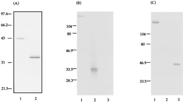
FIG. 2.
(A) Coomassie blue stain of SDS-PAGE of the recombinant CAT (lane 1) and GLU (lane 2) polypeptides. The CAT and GLU polypeptides were purified and resolubilized from inclusion bodies. Molecular weight markers in kilodaltons are indicated on the left. (B) Western blot of a crude extract of S. mutans GTFs (lane 1), purified GLU (lane 2), and purified CAT (lane 3). The blot was probed with biotinylated anti-GLU antibodies and developed with alkaline phosphatase-conjugated streptavidin. (C) Western blot identical to that shown in panel B but probed with biotinylated anti-CAT antibodies and developed with alkaline phosphatase-conjugated streptavidin.

