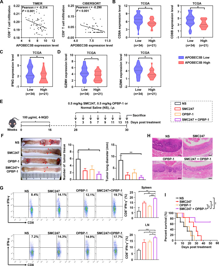
Figure 5.
The synergistic chemoprevention effects of 3, 5-diiodotyrosine combined with PD-1/PD-L1 blockade on 4-NQO induced ESCC mouse model. (A) CD8+ T cell infiltration was assessed based on mRNA expression level of APOBEC3B with TIMER and CIBERSORT. P values were determined by Wilcoxon test. (B–D) The mRNA expression of CD8A, CD8B and T effector genes were quantitatively analyzed between APOBEC3B low (n=34) and APOBEC3B high (n=21) expression status. *p<0.05 by one-tailed Student’s t-test. (E) Schematic illustration of the in vivo experiment. ESCC mice were i.p. injected with NS, 0.5 mg/kg 3, 5-diiodotyrosine, 0.5 mg/kg OPBP-1 or 3, 5-diiodotyrosine combined with OPBP-1 every 2 days for 2 weeks. (F) Tissues appearance of the esophageal epithelium, number and long diameter of tumor tissues in mice (n=3). (G) The proportion of IFN-γ+CD8+ T cells in spleen and lymph node cells was detected by intracellular cytokine staining (n=3, *p<0.05, **p<0.01, ***p<0.001). The data are presented as mean±SEM. (H) Histopathological assessment of esophagus tissues in each group was determined by H&E staining assay (scale bars: 100 µm). (I) Survival curve of mice (n=6).

