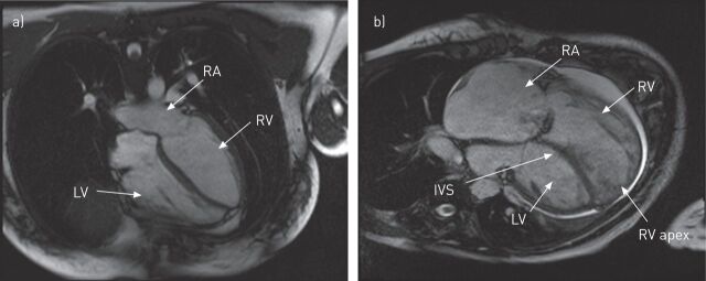
Figure 1.
Cardiac magnetic resonance imaging in patients with pulmonary hypertension. a) Patient showing right ventricular hypertrophy with normal sized right and left ventricular volumes. b) Patient with end-stage right ventricular failure and a severely dilated right ventricle and atrium. Where the septum is bulging to the left, the right ventricular apex is blunted. RV: right ventricle; LV: left ventricle; RA: right atrium; IVS: interventricular septum.

