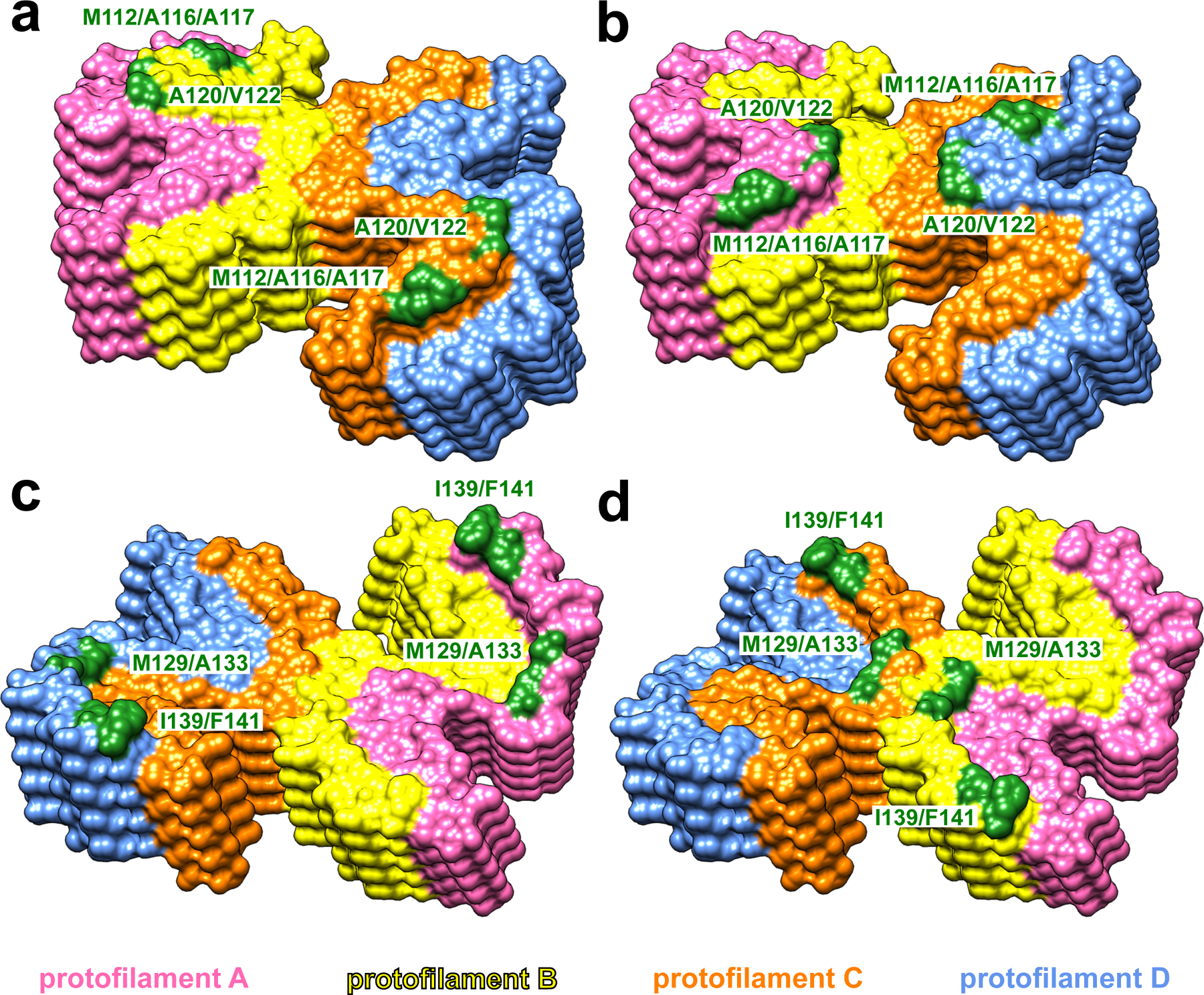
Fig. 2. Solvent-exposed amino acid residues at the top and bottom ends of huPrP23–144 fibrils.

At the top end of the fibril, two clusters of hydrophobic residues (M112/A116/A117 and A120/V122) in protofilaments B and C (yellow and orange, a) or protofilaments A and D (pink and blue, b) are exposed to the solvent. At the bottom end of the fibril, two clusters of hydrophobic residues (M129/A133 and I139/F141) in protofilaments A and D (pink and blue, c) or protofilaments B and C (yellow and orange, d) are exposed to the solvent.
