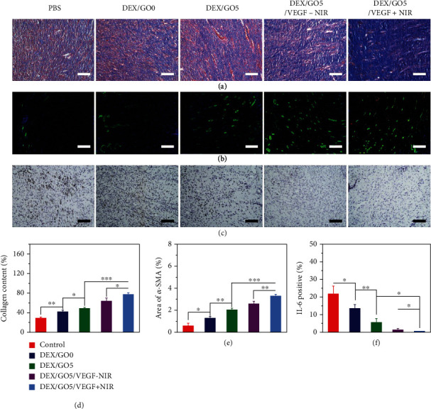
Figure 8.

Accelerated collagen deposition, promoted angiogenesis, and reduced inflammation. (a) Collagen deposition content after 11 days of wound healing. Scale bars: 100 μm. (b) Immunofluorescence assay of CD31 and α-SMA to indicating vascularization. Scale bars: 200 μm. (c) The immunohistochemistry of IL-6 of the granulation tissues for the different groups. Scale bars: 100 μm. (d) The collagen content (%) in the different groups calculated from Figure 7(a). (e) Statistical analysis of the relative coverage area of the α-SMA. (f) Statistical analysis of the IL-6 contents in the different groups.
