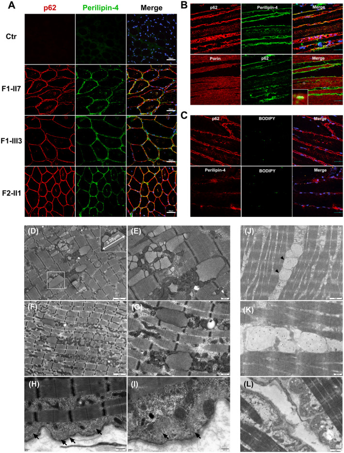
Figure 2.

Immunofluorescence, electron microscopy, and immunoelectron microscopy on muscle biopsy samples. (A and B) Co‐localization of perilipin‐4 and p62 in subsarcolemmal regions and within the cytoplasm of myofibers (A) and p62‐positive perilipin‐4 deposition (mean size 3.48 μm) distributed longitudinally within muscular fibers adjacent to porin‐positive mitochondria of F‐III3(B). (C) Colocalization of lipid droplets with p62 and perilipin‐4 in subsarcolemma and cytoplasm of myofibers in Patient S. (D) Vacuoles containing numerous membranous or myeloid bodies and (E–G) numerous membrane‐bound inclusions (mean size 3.08 μm) containing granulofilarmentous materials appeared between myofibrils under electron microscopy of Patient S (E) and F‐III3 (F and G) with clustered mitochondria around the inclusions. (H and I) Filamentous materials beneath basal membrane in Patient S (arrow). (J, K, and L) Immunofluorescence and immunoelectron microscopy (IEM) of Patient S revealed that the membrane‐bound inclusions and subsarcolemmal filamentous materials were p62 positive (arrowhead). Scale bars = 200 nm (I), 500 nm (E, G, H, and K), 1 μm (D and L), 2 μm (F and J), and 50 μm (A, B, and C).
