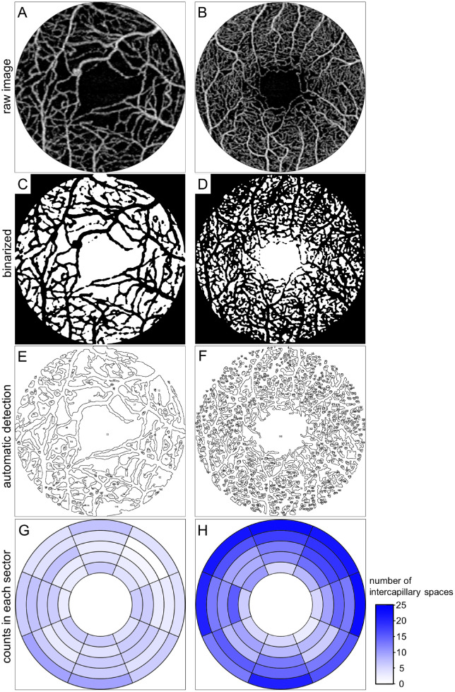
Figure 1.
Automatic quantification of intercapillary spaces in each sector on swept source OCTA images in two representative eyes with DR. (A, C, E, G) The decrease in the number of intercapillary spaces in a 42-year-old man with diabetic macular ischemia and PDR. (B, D, F, H) Abundant intercapillary spaces in a 52-year-old man with severe nonproliferative diabetic retinopathy (NPDR). (A, B) Raw OCTA images of the superficial slab within the central 2-mm circle. (C, D) Binary image (white = intercapillary spaces). (E, F) The intercapillary spaces are detected automatically. (G, H) Pseudocolor maps of the number of intercapillary spaces in each parafoveal sector.

