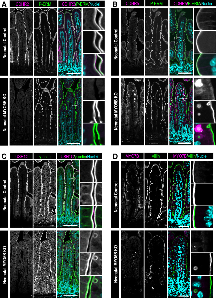
Figure 1.
Neonatal germline MYO5B KO mice exhibit altered IMAC gene expression and protein localization in the small intestine. Immunostaining of the small intestine for: the IMAC components (A) CDHR2 (magenta) and (B) CDHR5 (magenta), apical membrane marker phosphorylated-ezrin-radixin-moesin (P-ERM; green) and nuclei (blue) in neonatal control mice and germline MYO5B KO mice. Arrows indicate inclusions containing CDHR5 in the merged image. C: small intestinal tissue stained for the IMAC protein USH1C (magenta), the apical membrane protein γ actin (green) and nuclei (blue) in neonatal control and germline MYO5B KO mice. D: immunofluorescence images of the IMAC protein MYO7B (magenta), the apical membrane marker Villin (green) and nuclei (blue) in control and MYO5B KO mice. n = 4–6 mice/group. Scale bars = 50 µm. IMAC, intermicrovillar adhesion complex; KO, knockout.

