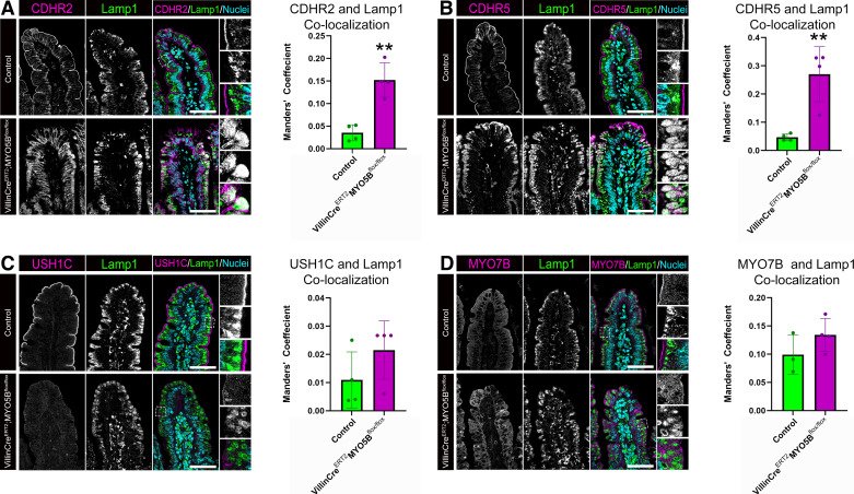
Figure 3.
Loss of MYO5B results in increased localization of CDHR2 and CDHR5 to Lamp1+ lysosomes. Adult control and VillinCreERT2;MYO5Bflox/flox mice were immunostained for the IMAC components (A) CDHR2 (magenta), (B) CDHR5 (magenta), (C) USH1C (magenta), (D) MYO7B (magenta), and Lamp1 (green) to identify lysosomes. Colocalization analysis of the degree of overlap between IMAC protein and Lamp1+ lysosomes was performed and plotted. **P < 0.01, n = 3 or 4 mice/group. Scale bars = 50 µm. IMAC, intermicrovillar adhesion complex.

