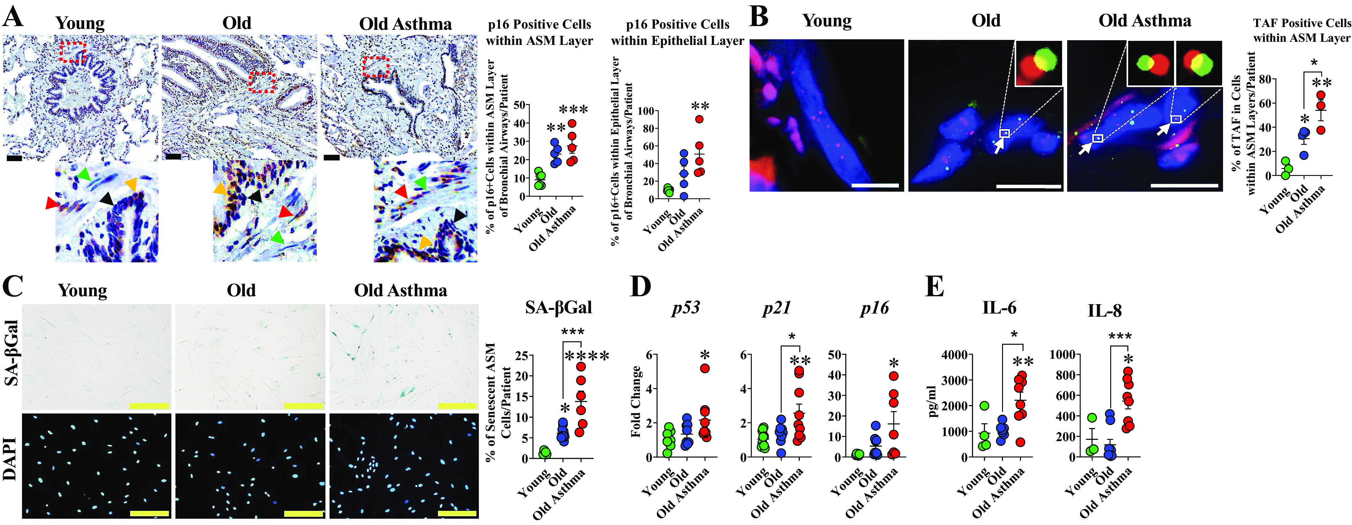
Figure 3.

Senescence in airway smooth muscle (ASM) cells in aging and asthma in the elderly (AIE). A: representative images of chromogenic staining for p16INK4A (dark brown) with cell nuclei counterstained with hematoxylin (blue). Red and orange arrows show p16INK4A positive cells while green and black arrows show p16INK4A negative cells within ASM and epithelial layers, respectively (n = 5 or 6 patients/group). Scale bar = 60 µm. B: representative images of quantitative fluorescence in situ hybridization (FISH) combined with immunofluorescence staining for γH2AX (red) and telomeres (green); cell nuclei counterstained with DAPI (blue). Accumulation of cell nuclei containing telomere-associated foci (TAF) within ASM layer was significantly higher in elderly asthmatic bronchial airway compared with elderly patients with nonasthma and young. White arrows point to TAF of colocalization at a single plane of Z-stack at higher magnification (top right, elderly and elderly asthma) (n = 3 or 4 patients/group). C: SA–βGal staining was increased in ASM cells from aging airways of elderly persons with asthma and elderly persons with nonasthma compared with young (n = 5–10 patients/group); Scale bar = 200 µm. D: qRT-PCR analysis showed significant changes in senescence-associated genes p53, p21, and p16 in ASM cells of elderly persons with asthma compared with young (n = 6–10 patients/group). E: ELISA of supernatants from cultured ASM cells of elderly persons with asthma showed increased IL-6 and IL-8 (n = 3–9). (*P < 0.05; **P < 0.01; ***P < 0.001; ****P < 0.0001). Means ± SE.
