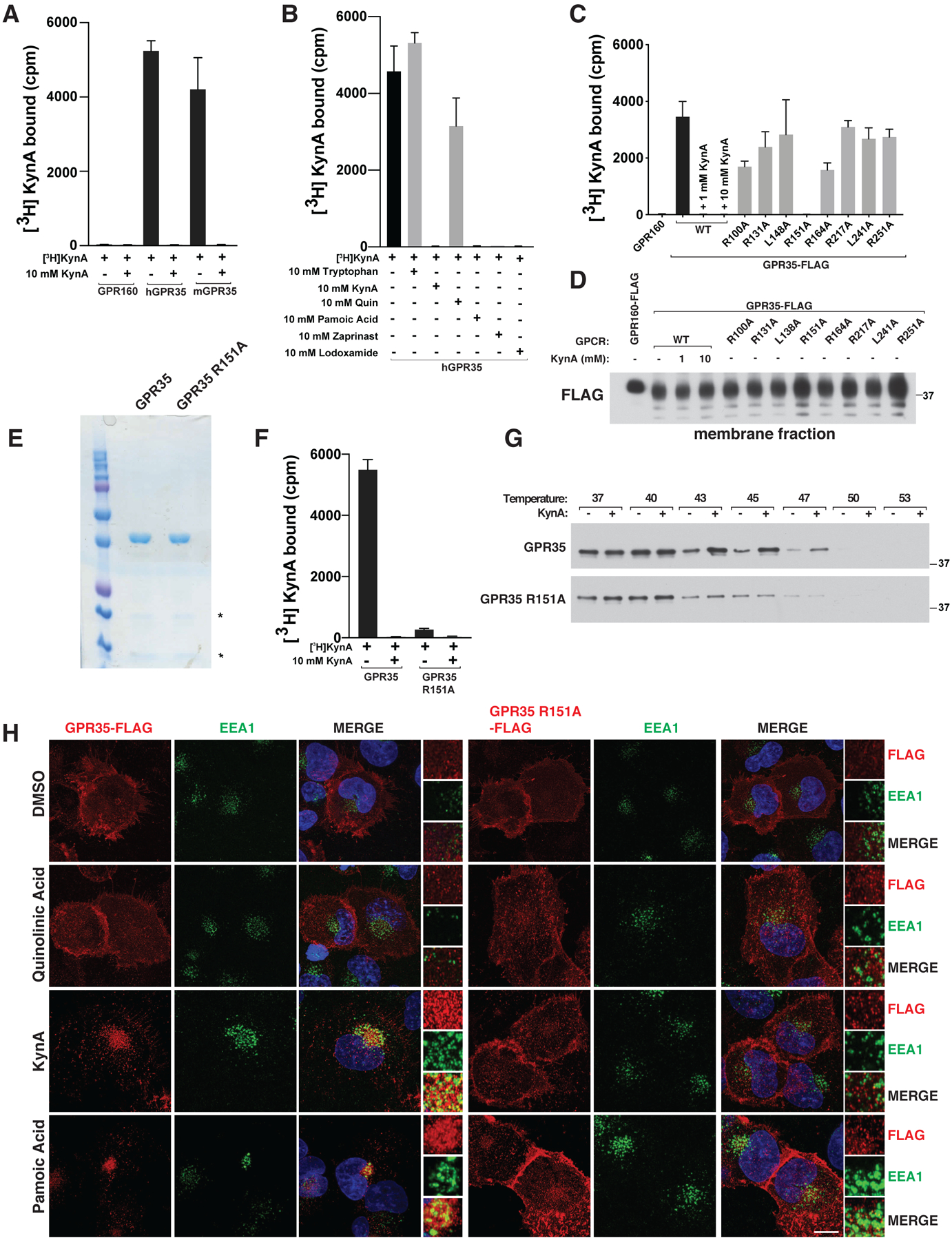
Figure 1: KynA binds GPR35 and promotes GPR35 internalization.

(A, B and F) [3H]-KynA binding assay using tandem affinity purified versions of the indicated HDL-reconstituted HA-FLAG-tagged GPCRs in the presence or absence of the indicated unlabeled competitor compounds (10 mM). (C and D) [3H]-KynA binding (C) and immunoblot (D) assays of isolated membrane fractions from 293T cells expressing FLAG-GPR160 or FLAG-GPR35 (wild-type or mutant). Where indicated, 1 and 10 mM unlabeled KynA was used as a competitor. (E and G) Commassie blue staining (E) and immunoblot (G) assay of the purified GPCRs used in (F). In (G) the proteins were pre-incubated with 1 mM KynA or DMSO for 1 hr at prior to exposure to the indicated temperature. (H) Confocal microscopy of mouse neonatal cardiomyocytes stably expressing wild-type or R151A GPR35-FLAG stimulated with 20 μM quinolinic acid, 20 μM KynA, or 1 μM pamoic Acid for 20 minutes. Scale bar indicates 20 μm. In (A), (B), (C), and (F), values are means −/+ SDs for three technical replicates from one representative experiment.
