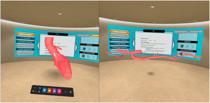
Figure 1:
Virtual room platform with all the clinical and image information of the patient on a panel and virtualized tridimensional model of the access route and landing zone. We can ‘walk around and inside’ the model analysing all anatomical details, the pattern of calcification and coronary ostia location. In addition, there is availability of tools to turn, expand, measure, draw, segment and visualize specific parts of the anatomy by enabling the transparency of tissues or take a pictures/videos and save them to the computer.

