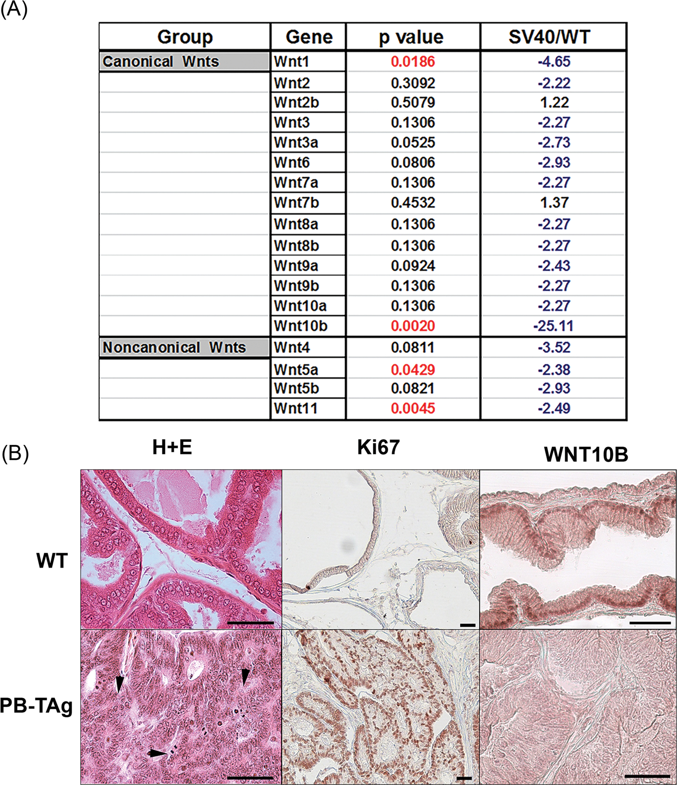
FIGURE 2.

A, Profile showing fold change in Wnt gene expression in probasin/TAg rat VPs at 25 weeks of age relative to WT prostates (SV40/WT) using a rat Wnt polymerase chain reaction array. Fold change in blue indicates a decrease and fold change in black is an increase relative expression levels. N = 3. B, Histology, Ki67 and WNT10B immunostaining in WT and probasin/Tag VPs at 25 weeks. (Top) WT VPs show normal glandular histology by hematoxylin and eosin stain, scarce proliferation by Ki67 labeling, and epithelial WNT10B immunostain. (Bottom) The probasin/Tag VP has widespread poorly differentiated adenocarcinoma (arrowheads), robust proliferation by Ki67 labeling, and marked reduction in epithelial WNT10B protein. Scale bar = 50 μM. VP, ventral prostate; WT, Wild type
