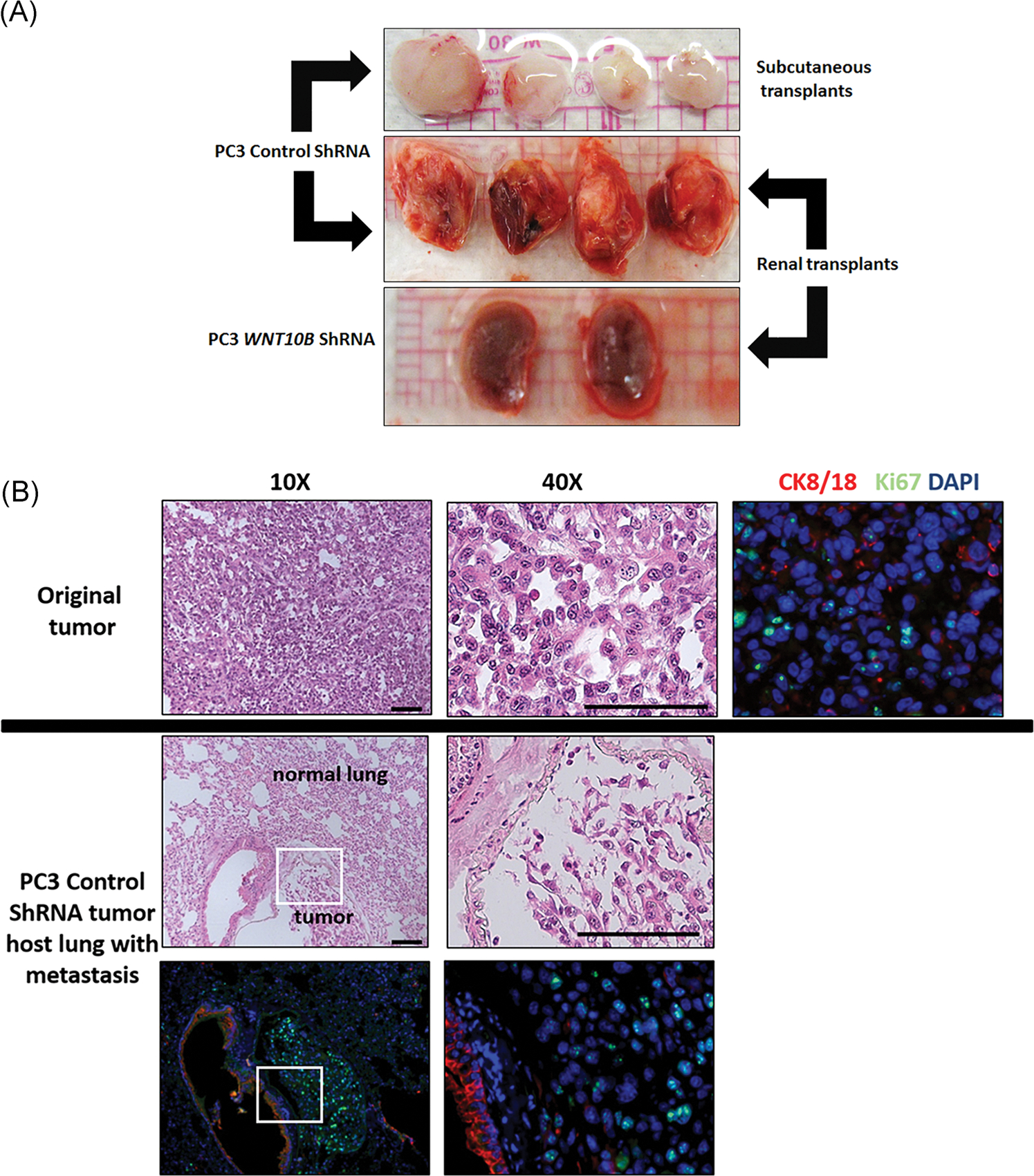
FIGURE 7.

A, Serial transplant of primary PC3 tumor with or without WNT10B knockdown. Primary tumors were allowed to form for 30 days using control shRNA or WNT10B shRNA-PC3 cells. Tumors were serially transplanted into nude mice hosts either s.c. or beneath the renal capsule and harvested after 9 weeks from s.c. sites (top) or renal site with kidney en masse (middle and bottom). Control shRNA-PC3 primary tumors formed new tumors at both sites (top and middle) whereas WNT10B shRNA-PC3 primary tumors were not transplantable at either site. The bottom panel shows kidneys grafted with WNT10B shRNA-PC3 primary tumors and only residual 1 mm grafts are visible. Images are representative of N = 8. B, Analysis of distant metastasis of PC3 cells in host mice. The top panel shows the primary s.c. tumor generated from control shRNA-PC3 cells with HE staining and immunohistochemistry for CK8/18 (red) and Ki67 (green) revealing poorly differentiated adenocarcinoma. The middle and bottom row images show lung tissue from host mice harboring control shRNA-PC3 metastasis. HE stain (middle) shows normal lung parenchyma with an infiltrated metastatic tumor at ×10 and ×40 magnification. CK8/18 (red) and Ki67 (green) immunostaining (bottom row) shows strong CK8/18 staining in lung epithelium with weaker CK8/18 and Ki67+ cells in the PC3 tumor metastasis. Bottom left is at ×10 and right is at ×40 of the tumor. DAPI, 4′,6-diamidino-2-phenylindole; HE, hematoxylin and eosin; s.c., subcutaneously; shRNA, short hairpin RNA
