
Fig. 3.
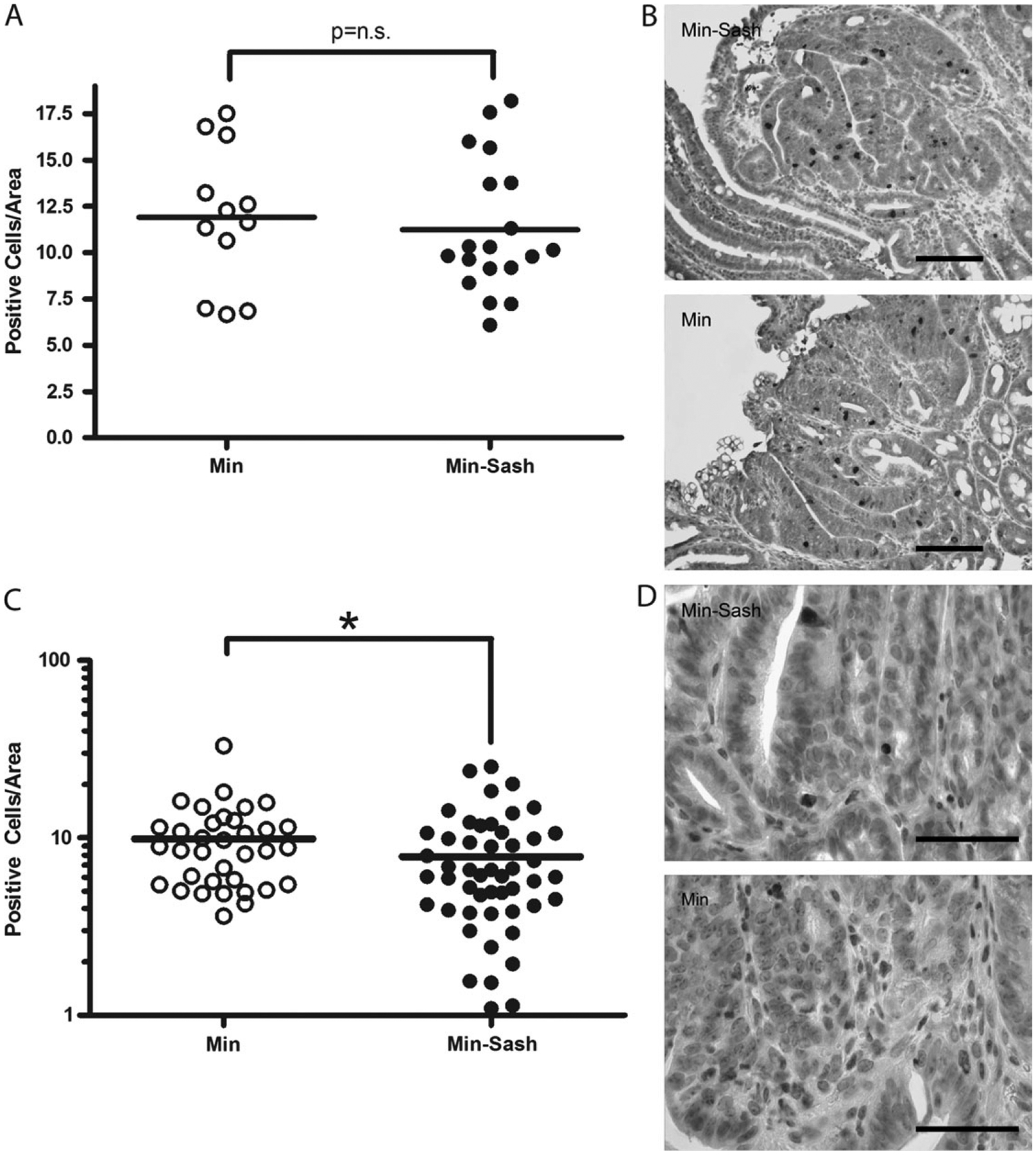
Apoptosis is inhibited in intestinal adenomas from Min–Sash mice compared with littermate controls; proliferation is not affected. (A) Intestinal adenomas were assayed for proliferative cells by phospho-histone H3 immunohistochemistry. Positive cells were counted per unit area using NIH ImageJ software; line, mean, not significant. (B) Phospho-histone H3 staining (dark brown staining) in tumors isolated from Min (top) and Min–Sash mice (bottom). Nuclei were visualized by counterstaining with hematoxylin. Scale bar indicates 100 μm. (C) Intestinal adenomas were isolated from Min (n = 7) and Min–Sash (n = 7) littermates and stained for cleaved caspase-3, a marker of apoptosis. Positive cells were counted per unit area as determined by NIH ImageJ software; line, mean, *P < 0.05, difference is statistically significant, Mann–Whitney two-tailed t-test. (D) High-power photomicrograph of caspase-3 immunohistochemistry (dark brown stain) in tumors isolated from Min (top) and Min–Sash (bottom) mice (×63), scale bar indicates 50 μm. Nuclei were counterstained with Mayer’s hematoxylin.