
Fig. 4.
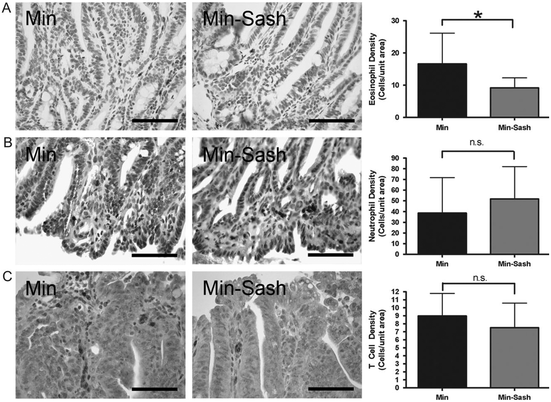
Eosinophils are less abundant in adenomas from Min–Sash mice compared with wild-type littermates; other leukocyte populations are not affected. Intestinal adenomas were isolated from wild-type and Min–Sash littermates and profiled for leukocyte populations by immunohistochemical staining. (A) Immunohistochemical demonstration of major basic protein-positive cells indicating eosinophils in wild-type (left) and Min–Sash (center) mice. Scale bar indicates 100 μm. Quantitation of eosinophils in Min–Sash mice compared with Min controls (right); *P < 0.05, difference is statistically significant, Student’s two-tailed t-test. (B and C) Immunohistochemical demonstration of neutrophils (anti-neutrophil+) (B) and T cells (CD3ϵ+) (C) in tumor tissue isolated from Min (left) and Min–Sash (center) mice. No differences were detected between groups (right). Scale bar indicates 100 μm.