Fig. 5.
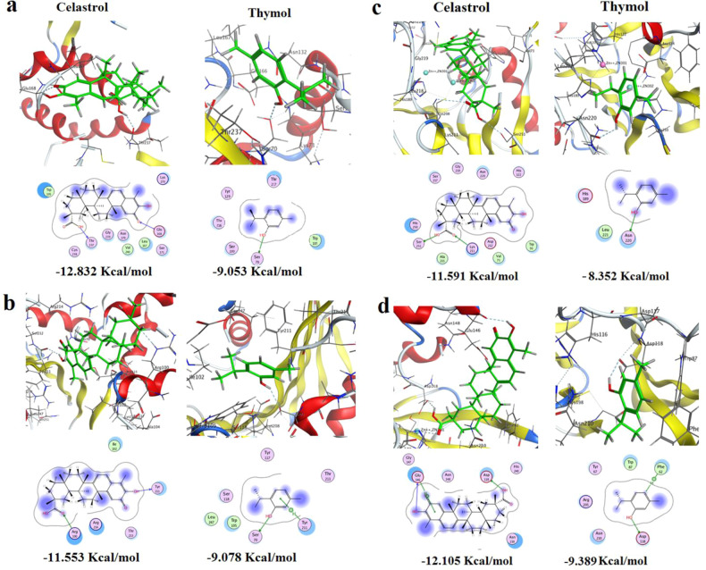
The putative binding modes (2D & 3D) of celastrol and thymol and their free binding energies expressed in Kcal/mol in the active site of the predicted 3D structure of K. pneumoniae carbapenemases a Class A KPC (KPC-2-3DW0), b Class D OXA (OXA-181 5OE0) c Class-B NDM (NDM-1 3SPU), d Class-B Verona Integron-encoded MBL (VIM-2 5YD7). The blue and cyan shadow of the ligand and active site amino acids respectively indicated strong hydrophobic/hydrophilic interactions

